Group: General Electric

Catalog excerpts

Stand out with SenoBright
Open the catalog to page 2
Know more. Now. Is something really there? What is it? Where is it? What’s next? For a woman facing even the slightest hint of a breast cancer diagnosis, the answers can’t come soon enough. , With SenoBright* you’ll have more information† to help you get to the answers you need. SenoBright is intended to be used as an adjunct following mammography and ultrasound exams to localize a known or suspected lesion.
Open the catalog to page 3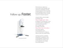
Performed as an adjunct to inconclusive mammography and ultrasound, SenoBright ContrastEnhanced Spectral Mammography (CESM) highlights areas of unusual blood flow patterns which may be cause for increased suspicion. Using an iodine contrast agent, SenoBright takes at different X-ray exposures. It then recombines them to highlight the contrast-enhanced areas— all in a simple, quick, procedure that takes less than ten minutes— much like a regular mammography exam. You’ll have answers to help you follow up with your patient faster. Senographe Essential Healthymagination-validated, SenoBright...
Open the catalog to page 4
Take a wait off your patients’ minds. With SenoBright, you can perform additional tests right away—using the same equipment, in the same room, with the same staff, on the same day. And help minimize the wonder and the worry of a concerned patient waiting for results.
Open the catalog to page 5
Four views can mean all the difference to a woman concerned about a breast cancer diagnosis. Simply SenoBright. One injection. One complete bi-lateral exam. Four views. The SenoBright acquisition is fully automated. The system automatically acquires the spectral data necessary to automatically create two images per view, a standard mammographic image showing tissue density, and a contrast-enhanced image in exactly the same position with the background signal subtracted out. With SenoBright, you stay in context. Whether you’re providing information to a breast surgeon, referring physician,...
Open the catalog to page 6
Solely SenoBright. A powerful X-ray tube and generator. A fast-reading digital detector. A proprietary recombination algorithm. With its unique ability to separate energy levels, GE technology advancements have made SenoBright possible. And GE offers the industry’s largest mammography field of view, on the Senographe Essential system.
Open the catalog to page 7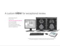
A custom view for exceptional review. Specifically designed to work with advanced applications such as SenoBright, the IDI Workstation— GE’s powerful reading tool— has the horsepower to handle advanced applications. Customized hanging protocols let each radiologist arrange images according to personal preference for exceptional reading efficiency—and see them the same, consistent way at every login.
Open the catalog to page 8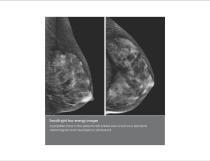
SenoBright low-energy images A palpable mass in the patient’s left breast was occult on a standard mammogram and visualized on ultrasound.
Open the catalog to page 10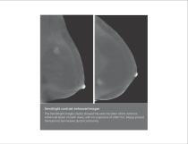
Standard mammography SenoBright contrast-enhanced images The SenoBright images clearly showed the exact location of the contrastenhanced lesion on both views, with no suspicions of other foci. Biopsy proved the lesion to be invasive ductal carcinoma.
Open the catalog to page 11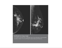
SenoBright low-energy images SenoBright CESM This patient presented with a palpable mass in her left breast. Mammography showed a cluster of microcalcifications and architectural distortion.
Open the catalog to page 13
SenoBright contrast-enhanced images The SenoBright images clearly showed the exact location of the contrastenhanced areas on both views, corresponding to the microcalcifications noted on the original mammography. Biopsy proved the lesion to be invasive ductal carcinoma.
Open the catalog to page 14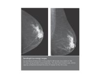
SenoBright low-energy images An architectural distortion in this patient’s left breast was visible on the CC view using standard mammography. She also had a known cyst, and its opacity somewhat blocked the area of interest on the MLO view.
Open the catalog to page 16
SenoBright contrast-enhanced images The SenoBright images clearly depicted the contrast agent and also subtracted out the cyst that was blocking the view in the standard mammograms. With the lesion well localized, the radiologist performed a biopsy, showing the lesion was invasive ductal carcinoma.
Open the catalog to page 17
Get to Yes or No sooner. In case after case… In patient after patient… SenoBright helps you get to Yes or No sooner— simply and smoothly, in a context you’re familiar with.
Open the catalog to page 18
©2012 General Electric Company – All rights reserved. General Electric Company reserves the right to make changes in specifications and features shown herein, or discontinue the product described at any time without notice or obligation. GE, GE Monogram, and imagination at work are trademarks of General Electric Company. *Trademark of General Electric Company. The photographs of women depicted in this brochure are not actual patients and the clinical cases do not correspond to these individuals. About GE Healthcare: Healthymagination is GE’s $6 billion commitment to bring high-quality...
Open the catalog to page 20All GE Healthcare catalogs and technical brochures
-
 Vscan Air™ CL
6 Pages
-
 Voluson™ Expert Series
4 Pages
-
 Vivid T9
9 Pages
-
 SIGNA™ Explorer
34 Pages
-
 Versana Active™
2 Pages
-
 GSI infographic
2 Pages
-
 MAC 2000 ECG Analysis System
5 Pages
-
 EMR Gateway Pro for MAC 2000
2 Pages
-
 Revolution EVO Gen 3
28 Pages
-
 Discovery MR750
5 Pages
-
 Seno Iris™
5 Pages
-
 SWAN
SWAN2 Pages
-
 GE Adventure Series™
14 Pages
-
 Mobile X-ray Systems
10 Pages
-
 GoldSeal
8 Pages
-
 Invenia™ABUS
6 Pages
-
 CardioMem CM 3000
2 Pages
-
 Lullaby Resus Plus
4 Pages
-
 Brivo NM615
5 Pages
-
 Aespire 7900
6 Pages
-
 Aespire View
4 Pages
-
 Aisys Carestation
12 Pages
-
 Avance Carestation
12 Pages
-
 Lunar iDXA Brochure
8 Pages
-
 Brochure Discovery IGS 730
9 Pages
-
 Prodigy for Bone Health
8 Pages
-
 Senographe Care
7 Pages
-
 SenoBright White Paper
7 Pages


