Group: LEO GROUP

Catalog excerpts

Full Digital PC-Based Ultrasonic Diagnostic APPLICABLE FIELDS » Suitable for the diagnosis ot Abdomen. Cardiac. Gynecology. Obstetrics, Thyroid Gland, Small Organs. Urology and so on. (Widely used tor clinical examination and diagnosis They are the ideal equipments to meet Hie needs ol various kinds of • Embedded computer platform is adopted in the ultrasonic mas- ■ DF £ Dynamic frequency scanning * Histogram. Sectionl drawing, Puncture fluids ■ Save hundreds of thousands of images and cine loop permanently • Dynamic feal-lime PiP local zoom functions ■ Double probe connectors • Multiple kinds or QB. measurement reports fetus physiological grades and reports and fetus growth curve • Auto-crsale report systems of Gyn., small cigars, cardiac, urol- ■ Compatible of jet printer, laser primer, video printer and: video IMAGE PROCESSING • Image process technologies: qontroiJabir* frame corre- lation, Gamma correction, edge enhancement, image smoothing, image denoisiong, auiomaiical gain ad- justment, up/down. left/right and black/white conver- ■ Dynamic Range. lOODb. 4 steps ot switches * Image magniiicaNon. stepless magnification, dynamic real-lime PIP local zoom functions ■ Cine loop. 256 frame auto/manual cine loop; niulti screens cine loop (4B, 9B); auto/manual cine loop un- • Image management system: Ihe functions ot pigeon- holing, browsing, comparing, saving, pnnlmg and lianslerring images; as many as hundreds ol thou- sands of images and thousands of cine loop could be saved; saved images could be operated by fuM- scieen browse under slide mode RUftMl+qwacy) rmitli-fiaqusncy] multi-frequency] Leading Digital Technologies DBF: Digital Beam Forming RDA: Real-time dynamic aperture imaging DRA: Dynamic real-time acoustic apodizer DRF: Dynamic receiving focus DFS: Dynamic frequency scanning, 4 kinds of scanning frequencies SOFTWARE FUNCTIONS_ • Measuremsm and calculation: measure perimeier and area by distance or ellipse method; measure perimeter and area by Liack method, measure body surlaoe area and volume by el- lipse method. A measure sticks; rate measure: linear stenosis ralio, area stenosis ratio, angpe measure. A31 calculations are • Assist tools: puncture puide. histogram, sectionl drawing Menu management interlace, reai time online support and: navigation clew system, image foreset and one-Key optimiza- tion functions • Auto-measure soltware of OB., Gyn.. small organs, cardiac. R US, OFD, THD, TIBIA. ULNA, FI, LIMP, BBT. FBP •Gyn.- uterus diameter, inlima Ifncknesv ovary colume, reg- nant ovarian toMicie, length ot cervix Jong^diarnetef, uterine • Small organs: thyioid gland, hip joinl • Cardiac: AOD, LAD, IVSTd, LVIDd. AA, LAD/AOD, LVPWd. LVIOs, EF, EF SLP. CA/CE, MVCF, CO. CI. LVMWl, AVSV. FS. • Urology: remained urine sample, proslate.PSAD • PresetMng system tor diagnosis and measurement formulas Dilferent formulas could be set according to different races. • Patienl cases database systems. Ah the data could be saved. • Multiple kinds of OB. measurement reports, fetus physiologi- cal grades ana repairs and Jetus growth cuive *Ajto-create report syslems ol Gyn., small organs, cardiac, ur- ology and oifter sections STANDARD CONFIGURATION_ • 10.4 inches SVGA high resolution n on-interlaces monitor • 256 frame auto/manual cine loop: rnulti screens cine loop [Ab. SB): auicimariual tine loop under B/fvt and M mode ■ 6.5MHz B:1ia transvaginal transducer (5.0-7,5fvlHz, mulli-tre- ■ 3.5MHz R15 cardiac transducer (2.5-5.DJvtHz, mulll-frequency} • Jet printer. Laser printer, Video printer and video lecorder RELATIVE EXTENDED PORTS_ • VGA, S-VideO, TV video port • USB2.0 port. 2G saving card • Multiple kinds of saving modes are all supported, containing hard disk, talsh disk. CF card. SD card and others. • Compatible of jet printer, laser printer, video printei and vid-
Open the catalog to page 1Archived catalogs
-
 LEO-3000H2 Ultrasound system
1 Pages
-
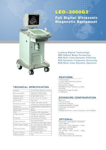 LEO-3000G2 Ultrasound system
1 Pages
-
 LEO-3000G1 Ultrasound system
1 Pages
-
 LEO-3000D2 Ultrasound system
1 Pages
-
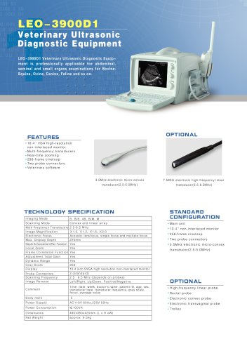
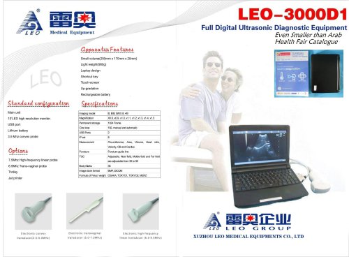
 LEO-3000D1 Ultrasound system
14 Pages
-
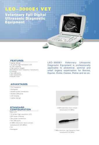
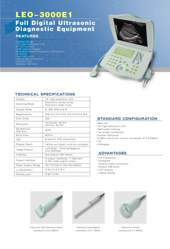 LEO-3000E1 Ultrasound system
1 Pages
-
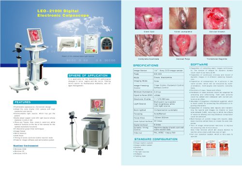 LEO-2100I colposcopy
1 Pages