Group: LEO GROUP

Catalog excerpts

•Suitable for the diagnosis ot Abdomen, Heart,Gynecology, Obstetrics.Thyroid Gland, Small Parts and so on. •Widely used for clinical examination and diagnosis. This is the ideal equipment to meet the needs of various kinds of hospitals and clinics. STANDARD CONFIGURATIONS • Main unit • 14 inch high resolution non-interlaced monitor • Images permanent storage •3.5MHz electronic convex transducer (2.5-5.0MH2) fllAIN UNIT Full digital trolley B/W diasonograph Sound beam processing: digital beam forming, continuously dynamic focusing, dynamic aperture Digital beam forming: AID conversion^ 10bil Double probe connectors More than 14 inches. Screen: Rotatable. progressive scanning without flicker Convex transducer (Abdomen): 2.5-5.0MHz High-frequency linear transducer (Small part): 6 5-8 5MHz Displaying depth: 240mm, and depth could be increased and stepless magnified Scanning modes: B. B/B. B/M, M. 4B (4B: the functions of real- time and freeze displaying provided) Focusing modes: dynamic-emitting focus, dynamic-receiving focus, dynamic aperture, adaptive acoustic lens Resolution: Horizontal: ^2mm Vertical: <1mm Geometrical precision: <5% Blind area: ^3mm 3.5MHz electronic convex transducer (2.5-5.0MHZ) Electronic transvaginal transducer (5.0-7.5MHZ) Electronic high-frequency linear transducer (6.5-8.5MHZ) The General Measurement and Analysis The Measuremenl and Analysis for Gynecology The Measuremenl and Analysis for Obstetrics The Measurement and Analysis for Urology The Measuremenl and Analysts for Small parts The Measuremenl and Analysis for Heart and the ambienl vessels neiumiu*. rui vach picture there are more than 5 parameters for reference, such as distance, area, perimeter, angle, volume, gestational age, fetus' weight, heart rate, slope rate, flux, left ventricle IMAGES PROCESS Planar images The functions of frame correlation, ycorrection and edge enhancement held, as well as the functions of pretreatment, post processing, left-right reversing and up-down reversing positive- negative reversing Image displaying ratio: stepless magnification which can be increased. Real-time local zooming held Maximum storage of more than 100.000 images. Images recorders Case history created and created automatically OTHER FUNCTIONS Output interface: AV, S-video. VGA. RS-232.RJ-45 and so on Jet printer, laser printer, video printer and video recorder optional DICOM 3.0 interface optional CD-ROM optional 8-section TGC adjustment, brightness adjustment and contrast Images management system. Case history database system and diagnosis & measurement pre-treat system Multi body marks Electronic high-lfequency Electronic convex
Open the catalog to page 1Archived catalogs
-
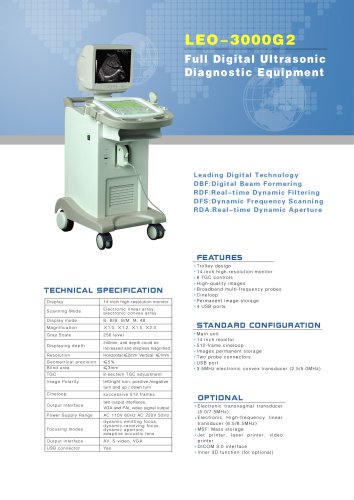 LEO-3000G2 Ultrasound system
1 Pages
-
 LEO-3000G1 Ultrasound system
1 Pages
-
 LEO-3000D2 Ultrasound system
1 Pages
-
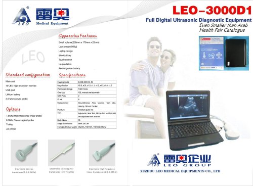
 LEO-3000D1 Ultrasound system
14 Pages
-
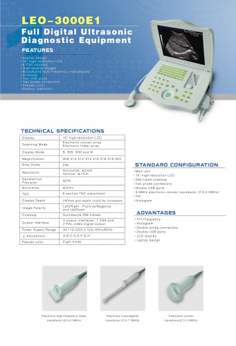 LEO-3000E1 Ultrasound system
1 Pages
-
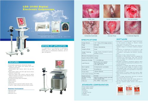 LEO-2100I colposcopy
1 Pages