
カタログの抜粋
rapifleX™ MALDI Tissuetyper™ For Mass Spectrometric Imaging Innovation with Integrity
カタログの1ページ目を開く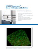
MALDI Tissuetyper™ Speed | Robustness | Definition The rapifleX instrument re-defines the key performance measures for MALDI Imaging: • Up to 50 true pixels / second for faster and better images • smartbeam™ 3D laser for better pixel-to-pixel reproducibility • Pixel size < 10µm for highest spatial resolution to retrieve biological information • Novel ion source design for increased robustness Lipids in rat testis, pixel size 10 µm, 395000 pixels, 200 shots per pixel, measurement time 165 minutes.
カタログの2ページ目を開く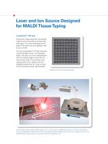
Laser and Ion Source Designed for MALDI Tissue Typing smartbeam™ 3D laser During the image acquisition the sample stage moves continually to achieve high pixel rates. The newly developed smartbeam™ 3D laser fires at a repetition rate of up to 10 kHz. The new smartbeam™ 3D laser features a laser diameter of 5µm. As illustrated below, the laser can move independently from the sample stage to scan the full area of each pixel. This produces truly square pixels which utilizes all of the available sample area for maximum sensitivity and pixel-to-pixel reproducibility. Ablated matrix showing...
カタログの3ページ目を開く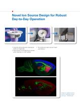
Novel Ion Source Design for Robust Day-to-Day Operation Extends uptime between cleaning by orders of magnitude. Easy access to self-aligning ion lenses when cleaning is in fact needed. The whole lens pack can be rinsed • with Ethanol. Proteins in sagittal rat brain section measured at 30 µm pixel size, ~150000 pixel.
カタログの4ページ目を開く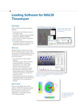
Leading Software for MALDI Tissuetyper flexImaging allows to set up and analyze individual datasets. • • • • • Wizard driven user-friendly set up of new experiments True 64-bit application allows efficient handling of megapixel datasets Direct access to single mass spectra Batch acquisition Co-register and super-impose virtual microscopic slides at full resolution for detailed histological annotation MALDI Imaging data overlaid with virtual microscopic slide in flexImaging SCiLS Lab statistical analysis software for mining MALDI Tissuetyper data. It allows both the analysis of individual...
カタログの5ページ目を開く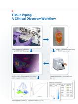
Tissue Typing - A Clinical Discovery Workflow Tissue samples are measured in the rapifleX MALDI Tissuetyper. MALDI Imaging dataset overlaid with micro- scopic image allows detailed annotation. Tissue is histologically stained after Tissue is scanned in a microscopic slide MzRange Pipeline-Aligned-Peafcs (ply Project) Centraid - Pipelin ± - Pipeline-Aligr p-Value - l-test Rating - t-test Statistical analysis of MALDI Tissuetyper data in SCiLS Lab.
カタログの6ページ目を開く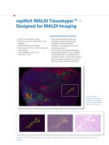
rapifleX MALDI Tissuetyper™ – Designed for MALDI Imaging Integrated software solutions • MALDI time-of-flight system • New ion source for robust day-to-day operation • Novel smartbeam™ 3D laser • Fast acquisition of up to 50 true square pixel / second • Laser repetition rate 10 kHz • Pixel size <10µm flexImaging dedicated molecular histology software controls data acquisition, data visualization, annotation and integration of virtual microscopic slides SCiLS Lab is the dedicated software for statistical analysis. From individual imaging datasets to complete clinical biomarker studies, it...
カタログの7ページ目を開く
rapifleX MALDI Tissuetyper™ – Customer Feedback “The ideal imaging MS system needs high data acquisition speeds, high spatial resolution capabilities, and high mass resolving power. With this new system, Bruker re-imagined the MALDI source and laser optics to deliver very impressive performance and information throughput. This is a revolutionary instrument which has the potential to significantly improve sample throughput and offer useful information in real time in clinical research.” Dr. Pierre Chaurand, Associate Professor, Université de Montréal, Canada “Translational clinically...
カタログの8ページ目を開くBruker Daltonics Inc./ブルカーのすべてのカタログと技術パンフレット
-
 IntelliSlidesTM
2 ページ
-
 CMC-assist
2 ページ
-
 Toxtyper
8 ページ
-
 Product Overview
16 ページ
-
 spotOn™
4 ページ
-
 Metabolomics
8 ページ
-
 TASQ Software
4 ページ
-
 timsTOF™
8 ページ
-
 TargetScreener
6 ページ
-
 timsTOF Pro
6 ページ
-
 Quant Proteomics
8 ページ
-
 MBT Galaxy RUO
4 ページ
-
 MBT Pilot RUO
4 ページ
-
 MBT SMART IVD
4 ページ
-
 MBT Consumables RUO
4 ページ
-
 MBT Sepsityper
6 ページ
-
 MBT BTS US
2 ページ
-
 MBT Pharma
8 ページ
-
 MALDI Imaging
16 ページ
-
 MALDI Biotyper CA System
12 ページ
-
 Bruker ToxScreener
6 ページ
-
 ProteinScape
8 ページ
-
 maxis II
12 ページ
-
 EVOQ
EVOQ6 ページ
-
 impact II
12 ページ
-
 micrOTOF II
8 ページ
-
 ImagePrep
4 ページ
-
 DE-tector
6 ページ
-
 pTD
pTD2 ページ
-
 RAID AFM
4 ページ
-
 RAID XP
4 ページ
-
 RAID S2
6 ページ
-
 RAID M100
6 ページ
-
 MM2
MM26 ページ
-
 SVG 2 and Probes
6 ページ
-
 µRAID
µRAID4 ページ
-
 VeroTect
4 ページ
-
 SIGIS II
2 ページ
-
 solarix XR
12 ページ
-
 The new autoflex speed
10 ページ
-
 Product Overview
20 ページ
-
 impact HD
12 ページ
-
 Toxtyper
6 ページ
-
 micrOTOF-Q III
6 ページ


カタログアーカイブ
-
 EVOQ - 2014
6 ページ