
カタログの抜粋

ORTHOPANTOMOGRAPH OP 2D OP 2D is a digital panoramic X-ray unit that combines distinctive design and reliable quality with all essential tools for standard panoramic imaging needs. OP 2D is part of the well-known ORTHOPANTOMOGRAPH™ product family. All the essentials included • Stable 5-point patient positioning • Optimal imaging geometry • Adjustable anterior layer position with 3 positioning lights • Powerful high-frequency X-ray tubehead design • High resolution CCD imaging sensor • V-shaped collimation optimizes image quality • Versatile software tools to enhance diagnostic ca
カタログの2ページ目を開く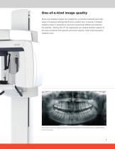
One-of-a-kind image quality Sharp and detailed images are enabled by a powerful tubehead and wide range of exposure settings that fit every patient size. A special V-shaped radiation beam is essential to overcome anatomical differences between the patients. Utilizing the OP 2D segmented pan feature enables capture of the area of interest with specific panoramic regions, while lowering patient radiation dose. The standard panoramic imaging program provides clear definition of the dental anatomy including TMJs— in only 10 seconds.
カタログの3ページ目を開く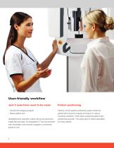
User-friendly workflow Just 2 selections need to be made Patient positioning • Choose the imaging program Intuitive 5-point patient positioning system holds the patient still during the imaging procedure to reduce movement artifacts. Three laser positioning lights make positioning accurate. The sharp layer is easily adjustable for every patient. • Select patient size Straightforward operation makes taking the panoramic image fast and easy. An integrated 5.7 inch touchscreen with remarkably user-friendly navigation is extremely simple to use.
カタログの4ページ目を開く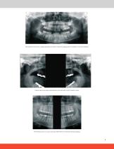
The pediatric panoramic imaging program provides a reduced imaging area for pediatric and small patients. Lateral view from temporomandibular joint area with mouth closed or open. The bitewing view is a quick and easy alternative for intraoral bitewing imaging.
カタログの5ページ目を開く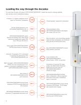
Leading the way through the decades For more than 60 years, the name of ORTHOPANTOMOGRAPH™ system has stood for ultimate reliability and clinically correct maxillofacial imaging. Professor Y.V. Paatero publishes his first paper on Panoramic Tomography. The first dental panoramic x-ray, ORTHOPANTOMOGRAPH OP1, is developed. ORTHOPANTOMOGRAPH system becomes the leading name within dental panoramic imaging with models OP5/ OC5, OP6 and OP10/OC10. Direct digital ORTHOPANTOMOGRAPH OP100 product family is introduced. “Pantomography” equipment is presented. New ORTHOPANTOMOGRAPH product family...
カタログの6ページ目を開く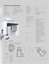
Technical specifications Panoramic Image Detector Sensor Pixel Size Image Pixel Size current limited with 100 25 VAC) -1 Image Field Height Nominal Voltage Exposure Time Imaging Programs Standard, Pediatric, Segmented, TMJ Minimum Total Filtration Wheelchair accessible Focal Spot Tube Voltage Tube Current Lateral, Bitewing Weight Minimum System Requirements for Acquisition Workstation CPU (processor) Intel Core i3, 2-cores or more GPU (graphics processing unit) Storage (hard disk) 8 GB free disk space, 100 GB or more recommended, plus backup Gigabit Ethernet 1000 Mb/s or Fast Ethernet...
カタログの7ページ目を開く
About DEXIS DEXIS innovation is nothing new. After all, our recognized, trusted products are built on over 200 years of dental imaging expertise, combining leading brands including i-CAT™, Gendex™, Instrumentarium, SOREDEX™, and NOMAD™ Pro 2. Today, over 150,000 offices trust DEXIS products around the world. DEXIS now includes a full portfolio of products including CBCT and Intraoral scanners, our legacy digital sensors and handheld x-ray system and DTX Studio™ Clinic, the next generation software. This complete digital solution works seamlessly together as well as with other systems to...
カタログの8ページ目を開くDEXISのすべてのカタログと技術パンフレット
-
 OP 3D
OP 3D9 ページ
-
 IXS
IXS4 ページ
-
 Nomad Pro 2
8 ページ
-
 DEXISTM Titanium by KaVo
12 ページ