
カタログの抜粋
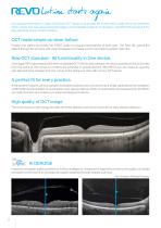
lution starts again Our supreme experience in Spectral Domain OCT allows us to provide the market with a state of the art instrument which comes with new advanced technologies and remarkable simplicity of operation. The REVO 80 will expand the daily demands of any modern practice. OCT made simple as never before Position the patient and press the START button to acquire examinations of both eyes. The Revo 80, guides the patient through the process with vocal messages to increase comfort and reduce patient chair time. New OCT standard - All functionality In One device. Once again REVO goes...
カタログの2ページ目を開く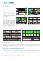
A single 3D Retina scan performs both Retina and Glaucoma analysis. The software automatically recognizes 8 retinal layers which assists with precise diagnosis and mapping of any changes in the patient’s condition. A variety of result analysis and presentation methods allows the most suitable selection to increase efficiency. REVO’s standard high density scanning capability and blood vessel structure recognition enable a precise alignment of past and current scans. The operator can analyse changes in morphology, quantified progression maps and evaluate the progression trends. Progression...
カタログの3ページ目を開く
lution starts again This non-invasive dye free technique allows the visualization of the microvasculature of the retina. Both blood flow and structural visualization give additional diagnostic information about many retinal diseases. Angiography scan allows assessment of the structural vasculature of the macula, the periphery or the optic disc. Extremely short scanning times of 1,6 seconds in standard resolution or 3 seconds in high resolution. Now Angiography OCT can become a routine in your diagnostic practice. The Angiography mosaic delivers high-detail images over a large field of the...
カタログの4ページ目を開く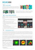
Comprehensive glaucoma analytical tools for quantication of the Nerve Fiber Layer, Ganglion layer and Optic Head with DDLS enable the user to perform precise diagnosis and monitoring of glaucoma over time. With the Asymmetry Analysis of Ganglion layers between hemispheres and between eyes it is possible to identify and detect glaucoma in early stages and in non-typical patients. Invaluable combination of information about the functional quality of vision with comprehensive data on retinal Ganglion Cells, RNFL and Optic Nerve Head for both eyes on a single report page. The S&F report...
カタログの5ページ目を開く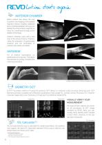
lution starts again ANTERIOR CHAMBER Built-in anterior lens allows the user to perform the imaging of the anterior segment without installing additional lens or forehead adapter. Now you can display the whole anterior segment or focus on a small area to bring out the details of the image. Anterior Chamber exam with a fast view of the whole Anterior Chamber make the evaluation of gonioscopy situation and the verification of cataract lens easier and faster. For all anterior examination, no additional lens is required. This allows the examiner to quickly complete the scanning procedure. *...
カタログの6ページ目を開く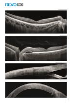
Choroidal observation Wide Central scan Sclera and Anterior Structure Cornea wide scan
カタログの7ページ目を開く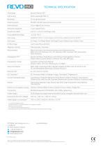
TECHNICAL SPECIFICATION Technology Light source Scanning speed Axial resolution Transverse resolution Overall scan depth Focus adjustment range Scan range 80 0005 / 60 000 measurements per second 2.6 μm digital, 5 μm in tissue 12 μm, typical 18 μm 2.8 mm / ~6 mm in Full Range mode -25 D to +25 D Posterior 5 mm to 15 mm, Angio 3 mm to 6 mm, Anterior 3 mm to 18 mm 3D, Angio1, Full Range Radial, Full Range B-scan, Radial, B-scan, Raster, Cross Scan types Fundus image Live Fundus Reconstruction Retina analysis Retina thickness, Inner Retinal thickness, Outer Retinal thickness, RNFL+GCL+IPL...
カタログの8ページ目を開くOptopol Technologyのすべてのカタログと技術パンフレット
-
 REVO 60
8 ページ
-
 REVO FC 130
16 ページ
-
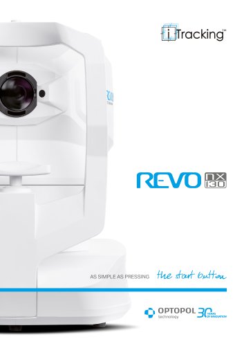 REVO NX 130
12 ページ
カタログアーカイブ
-
 revo-fc130
16 ページ
-
 REVO NX
12 ページ
-
 REVO NX 130
12 ページ
-
 REVO FC
6 ページ
-
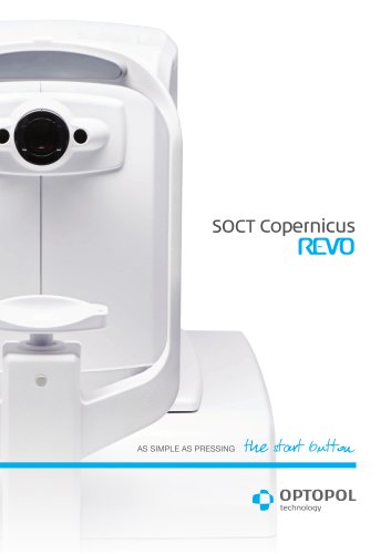 SOCT Coperncius REVO
6 ページ
