カタログの抜粋

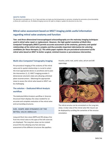
Mitral Valve The pre-op assessment tool for Mitral valve repair and replacement Determine the anatomy and dimensions of the patient’s Mitral valve with this dedicated workflow. The 3D shape and dimensions of the annulus can easily be defined as well as the relationship with surrounding structures. Assess different approach routes to get a complete overview of the patient’s anatomy. The heart with a defined Mitral annulus and a virtual valve Mitral annulus anatomy Mitral annulus Saddle shape and D-shape Annulus annotation A single click brings you into the Mitral space. Choose for an automatic trace of the saddle shaped annulus or use an easy manual method. The dimensions of the Mitral annulus are automatically calculated. A D-shape model is also available. Anatomical assessment Different views are available to assess the shape and position of calcium and vessel centerlines can be traced and visualized. Annulus dimensions Multimodality mitral assessment Optimal projection The simulated Angio view can be used to find optimal projections in order to save time during the procedure. 3D echo with virtual valve and neo-LVOT A virtual valve can be placed, and a neoLVOT measurement can be done directly on 3D echo measurements. The 3D echo data can be linked with the CT-analysis for a better assessment. After tracing the annuli within both modalities, the data can be linked, combining the best of both worlds. Philipsweg 1 6227 AJ Maastricht The Netherlands Simulated Angio view
カタログの1ページ目を開く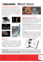
Mitral Valve Virtual valve and neo-LVOT on CT A virtual valve can be placed by means of importing an STL file or creating a custom valve. After a virtual valve is placed the LVOT obstruction can be measured and saved to the report both in end-systole and end-diastole. Left: Virtual valve Right: Short axis view on the LVOT Approach route assessment Septal Crossing Septal Crossing: This workflow can be used to plan and visualize the catheter path through the Vena Cava and Fossa Ovalis by determining the septum anatomy. Virtual TEE and ICE: Make your pre-op planning complete with the virtual...
カタログの2ページ目を開くPie Medical Imagingのすべてのカタログと技術パンフレット
-
 CAAS QVA
2 ページ
-
 CAAS QCA brochure
2 ページ
-
 CAAS LVA
2 ページ
-
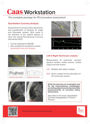
 CAAS Workstation
2 ページ
-
 RVA
RVA2 ページ
-
 Stent Enhancer
2 ページ
-
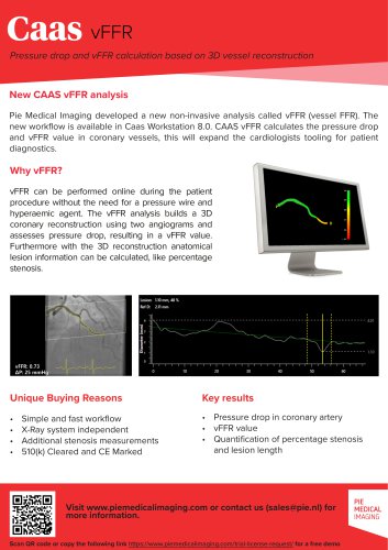 CAAS vFFR
2 ページ
-
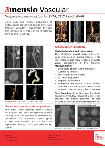 3mensio vascular
2 ページ
-
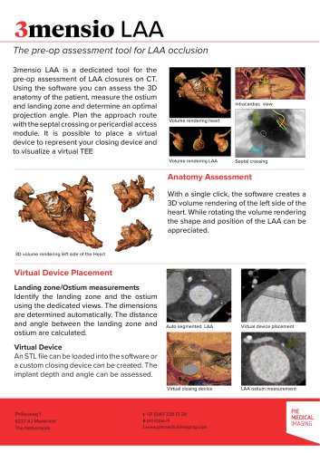 3mensio LAA
2 ページ
-
 3mensio Aortic valve
2 ページ
-
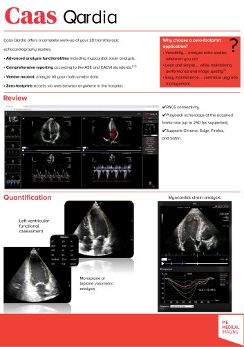 Caas Qardia
2 ページ
-
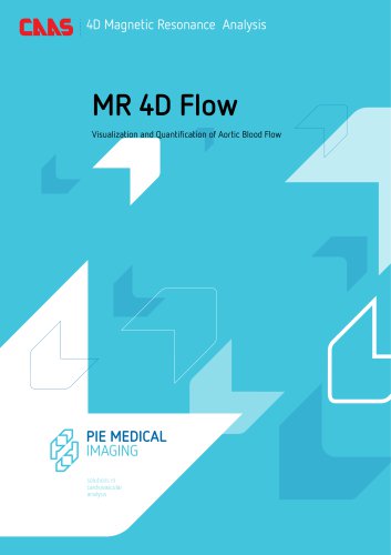
 MR-4D-Flow
2 ページ
-
 CAAS-IntraVascular
2 ページ
-
 Caas IV-LINQ
2 ページ
-
 StentEnhancer
4 ページ
-
 MRV | MR Flow
6 ページ
-
 QCA 3D
5 ページ
-
 CAAS MRV
6 ページ
-
 CAAS QCA whitepaper
5 ページ

カタログアーカイブ
-
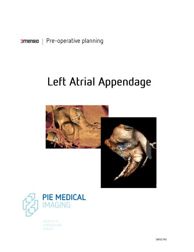
 LAA
LAA6 ページ
-
 MR 4D Flow
6 ページ
-
 CAAS IntraVascular
6 ページ
-
 3mensio Aortic Root
6 ページ