- Company
- Products
- Catalogs
- News & Trends
- Exhibitions
C41
1 /16Pages

C41
1 /16Pages
Catalog excerpts

…going one step further
Open the catalog to page 1
Fila radicularia Radix posterior Ganglion sensorium nervi spinalis Truncus nervi spinalis R. posterior R. anterior R. communicans R. meningeus Radix anterior Lig. denticulatum Dura mater spinalis Arachnoidea mater spinalis Spatium subarachnoideum Pia mater spinalis A. spinalis anterior Vv. spinales anteriores Substantia grisea Substantia alba Cornu posterius Cornu anterius Commissura grisea Sulcus posterolateralis Sulcus intermedius posterior Sulcus medianus posterior Fissura mediana anterior Commissura alba Canalis centralis Sulcus anterolateralis Septum medianum posterius Pars cervicalis Pars...
Open the catalog to page 3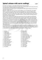
Spinal column with nerve endings The model is an enlarged 5:1 scale model of how the spinal cord is constructed. The spinal cord is composed from both grey and white matter. The grey matter is on the inside and encloses the central channel. The white matter faces towards the outside. The surface of the spinal cord has a groove running down the middle along the length of the front-facing side (fissura median anterior). Beside this, another fissure can be seen also running lengthways (sulcus anterolateralis). Some shallow fissures can also be seen on the surface of the spinal cord facing the rear....
Open the catalog to page 4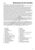
Das Modell zeigt in ca. 5-facher Vergrößerung den Aufbau des Rückenmarks. Das Rückenmark setzt sich aus der grauen und weißen Substanz zusammen. Die graue Substanz liegt innen und umschließt den Zentralkanal. Die weiße Substanz befindet sich außen. Die Oberfläche des Rückenmarks zeigt auf der vorderen Seite in der Mitte eine längsverlaufende Spalte (Fissura mediana anterior). Seitlich davon ist jeweils wiederum eine längsverlaufende Furche (Sulcus anterolateralis) erkennbar. Auf der nach hinten gerichteten Oberfläche des Rückenmarks sind flache Furchen sichtbar: Mittig liegt der Sulcus medianus...
Open the catalog to page 5
Médula espinal con terminaciones nerviosas Español El modelo representa la estructura de la médula espinal aumentada aproximadamente 5 veces de tamaño. La médula espinal se compone de substancia gris y substancia blanca. La substancia gris se encuentra en la parte interna y encierra el conducto central. La substancia blanca esta ubicada en la parte externa. La superficie de la médula muestra en su parte anterior, en la mitad, una hendidura longitudinal: la fisura mediana anterior. A cada lado de ésta, se reconoce un surco anterior lateral (Sulcus anterolateralis). En la superficie posterior de...
Open the catalog to page 6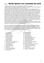
Moelle épinière avec extrémités des nerfs Le modèle représente la structure de la moelle épinière agrandie environ cinq fois. La moelle épinière est constitué de substance grise et blanche. La substance grise est située à l’intérieur et entoure le canal central. La substance blanche se situe à l’extérieur. La surface de la moelle épinière présente au milieu de sa face avant une fissure longitudinale (Fissura mediana anterior). Sur le côté, on découvre un autre sillon longitudinal (Sulcus anterolateralis). Des sillons plats sont visibles à la surface arrière de la moelle épinière. Au centre se...
Open the catalog to page 7
Medula espinhal com terminações nervosas O modelo, aumentado aproximadamente 5 vezes, mostra a estrutura da medula espinhal. A medula é formada pela substância cinza e a substância branca juntas. A substância cinzenta encontra-se na parte interna e cobre o canal central. A substância branca encontra-se na parte exterior. A superfície da medula apresenta no lado frontal, medianamente, uma prega de percurso longitudinal (Fissura mediana anterior). A cada lado desta, reconhece-se um sulco de percurso longitudinal (Sulcus anterolateralis). Na superfície orientada para trás, são visíveis sulcos planos....
Open the catalog to page 12
Midollo spinale con terminazioni nervose Il modello, ingrandito di ca. 5 volte, mostra la struttura del midollo spinale. Il midollo spinale è composto dalla sostanza grigia e dalla sostanza bianca. La sostanza grigia si trova all’interno e circonda il canale centrale. La sostanza bianca si trova all’esterno. La superficie del midollo spinale mostra al centro della parte anteriore una fessura longitudinale (Fissura mediana anterior). Invece, lateralmente si può riconoscere un solco anterolaterale (Sulcus anterolateralis). Sulla superficie del midollo spinale rivolta all’indietro sono visibili...
Open the catalog to page 13
脊髄の構造がよくわかる5倍大サイズのモデルです。 脊髄は灰白質と白質とで構成されており,灰白質は中心管の周りを囲むように内側に,白質は外側に位置して います。脊髄表面の前側中央には縦方向に走る溝(前正中裂)があります。対して,後側の中央にも縦方向 の長い溝(後正中溝)があり,その両側に浅い溝(後外側溝)が走っています。頚部と上部胸椎部にはもう一 つ,中間溝(後中間溝)があります。また後正中溝からは脊髄内部に向けて後正中中隔が形成されています。 根糸と呼ばれる神経の集まりは,脊髄の前側から後側へと続き,前根と後根を形作ります。脊髄神経節は前根 との接合部
Open the catalog to page 14
3B Scientific 3B Scientific GmbH Rudorffweg 8 • 21031 Hamburg • Germany Tel.: + 49-40-73966-0 • Fax: + 49-40-73966-100 www.3bscientific.com • [email protected] © Copyright 2005 / 2014 for instruction manual and design of product: 3B Scientific GmbH, Germany
Open the catalog to page 16All 3B Scientific catalogs and technical brochures
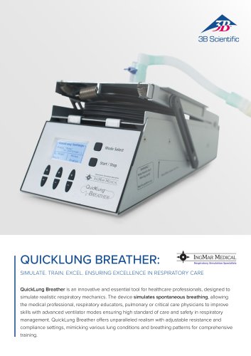
 QuickLung Breather
12 Pages
 SAM4 - Auscultation Manikin
9 Pages
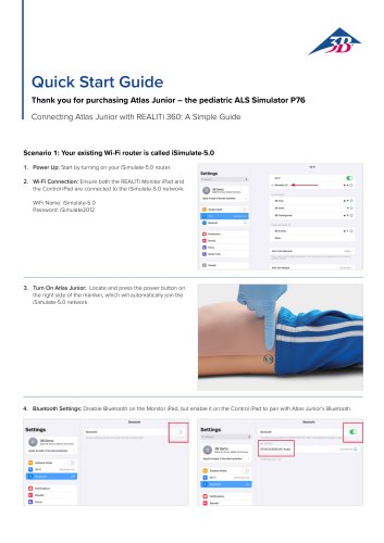
 Quick Start Guide Atlas Baby
4 Pages
 Quick Start Guide Atlas
4 Pages
 Sellsheet eSono Abdominal
3 Pages
 Sellsheet eSono MSK
3 Pages
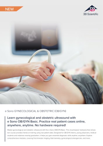 Sellsheet eSono OBGYN
3 Pages
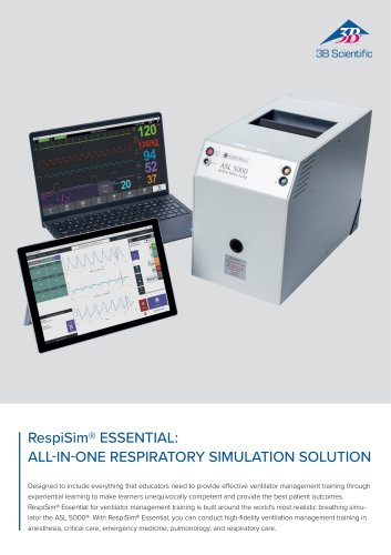 Sellsheet IngMar RespiSim
2 Pages
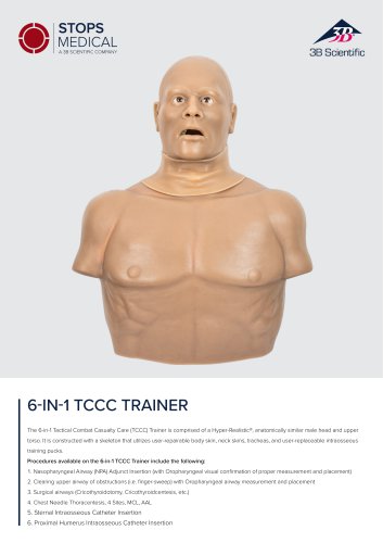 Sellsheet Stops 6N1 Trainer
2 Pages
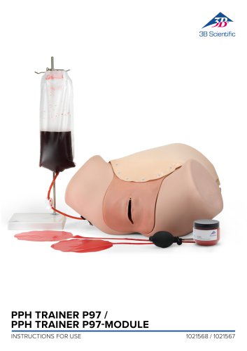 PPH Trainer P97
14 Pages
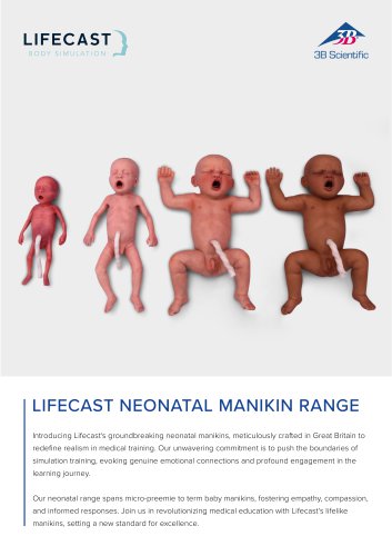 Sellsheet Lifecast Neonatal
3 Pages
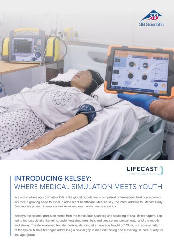 Sellsheet Lifecast Teenager
2 Pages
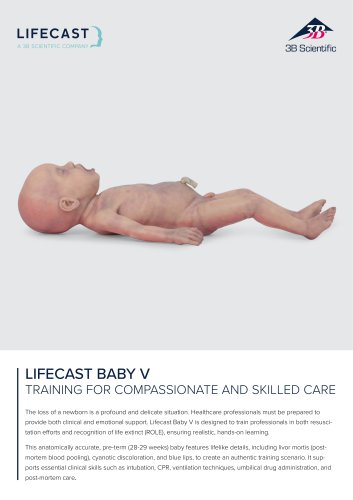 Sellsheet Lifecast Baby V
2 Pages
 Product Manual Atlas Baby
16 Pages
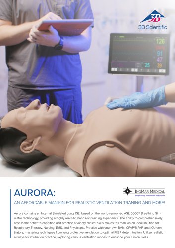 IngMar Sellsheet Aurora
2 Pages
 IngMar Sellsheet QuickLung
2 Pages
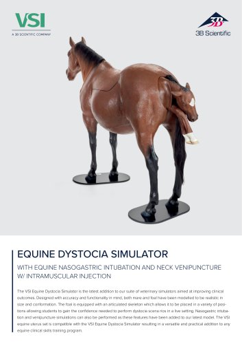 Sellsheet VSI 1025586
2 Pages
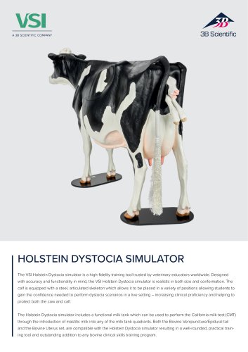 Sellsheet VSI 1025528
2 Pages
 Sellsheet VSI 1025662
2 Pages
 Sellsheet VSI 1025616
2 Pages
 Immersive Brochure
13 Pages
 Medical Simulation
51 Pages
 Lifecast Brochure
16 Pages
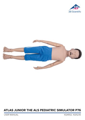
 Atlas Product Manual
18 Pages
 Cardionics Brochure Simulation
23 Pages
 Acupuncture
35 Pages
 Best of Therapy
12 Pages
 Manual P120/P121/P122/P124/P125
60 Pages
 P10/1,P11/1 product manual
11 Pages
 P16 Product brochure
2 Pages
 Medical Simulation EMS TCCC
9 Pages
 Catalog Natural Sciences
196 Pages
 K17
K1716 Pages
 D25 Half Lower Jaw
13 Pages
 D20 Dentition Development
12 Pages
 D10
D1012 Pages
 L56
L5630 Pages
 L50, L51, L55
36 Pages
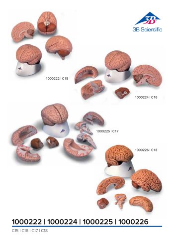
 C15, C16, C17, C18, C20
12 Pages
 P80 SIMone Product Manual
52 Pages
 P57 Quick instructions
16 Pages
 N30 / N31 Product Manual
12 Pages
 N15 Acupuncture Ears
2 Pages
 P16 Product manual
8 Pages
 P10CCD product brochure
2 Pages
 P10CCD product manual
16 Pages
 P72+light Product manual
28 Pages
 P72+light Product brochure
2 Pages
 Female Breast
30 Pages
 C18
C189 Pages
 G01
G0124 Pages
 3B Smart Anatomy
3 Pages
 M10
M1016 Pages
 A291
A29120 Pages
 F11
F1113 Pages
 P72
P7248 Pages
 B60
B6016 Pages
 A05/2 ,A11, A13
18 Pages
 A290 A291
20 Pages
 G21, G22
9 Pages
 K25
K2512 Pages
 K20, K21
12 Pages


![Product Manual - I.v. Injection Arm P50/1 - P50/1 [1021418]](https://img.medicalexpo.com/pdf/repository_me/67454/product-manual-iv-injection-arm-p50-1-p50-1-1021418-249392_1mg.jpg)

![Product Manual - Hemorrhage Control Arm Trainer P102 - P102 [1022652]](https://img.medicalexpo.com/pdf/repository_me/67454/product-manual-hemorrhage-control-arm-trainer-p102-p102-1022652-249356_1mg.jpg)
![Product Manual - Trainer for wound care and bandaging techniques - P100 [1020592]](https://img.medicalexpo.com/pdf/repository_me/67454/product-manual-trainer-wound-care-bandaging-techniques-p100-1020592-249350_1mg.jpg)
![Product Manual - Postpartum Hemorrhage Trainer - PPH Trainer P97 - P97 [1021568]](https://img.medicalexpo.com/pdf/repository_me/67454/product-manual-postpartum-hemorrhage-trainer-pph-trainer-p97-p97-1021568-249337_1mg.jpg)


- Hosley anatomical model
- Hosley training anatomical model
- Hosley teaching anatomical model
- Hosley training simulator
- Hosley general care simulator
- Hosley tooth model
- Hosley upper body simulator
- Hosley flexible anatomical model
- Hosley bone model
- Dental care model
- Hosley skull model
- Hosley training manikin
- Hosley surgery simulator
- Stethoscope
- Hosley plastic anatomical model
- Hosley mouth anatomical model
- White anatomical model
- Patient simulation unit
- Vascular model
- Training vascular model