BioCAM X brochure
BioCAM X brochure
Catalog excerpts

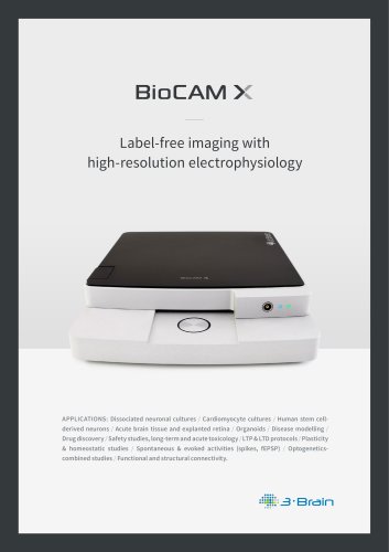
Label-free imaging with high-resolution electrophysiology APPLICATIONS: Dissociated neuronal cultures / Cardiomyocyte cultures / Human stem cellderived neurons / Acute brain tissue and explanted retina / Organoids / Disease modelling / Drug discovery / Safety studies, long-term and acute toxicology / LTP & LTD protocols / Plasticity & homeostatic studies / Spontaneous & evoked activities (spikes, fEPSP) / Optogeneticscombined studies / Functional and structural connectivity.
Open the catalog to page 1
Originating from 3Brain’s expertise gained in the manufacture of the first CMOS high-resolution multielectrode arrays, BioCAM X will boost your research capabilities by enabling simultaneous recordings from a total of 4096 electrodes sampled at 18 kHz per electrode. You can either choose to store the entire raw signals captured by the BioCAM X or to take advantage of the several available degrees of compression, which will allow you to save space on your hard disk and thus decrease the computational resources required for further data processing. All-in-one BioCAM X incorporates further optional...
Open the catalog to page 2
Whatever your experimental needs with multi-electrode arrays are, BioCAM X can satisfy them! Its high sampling frequency and a user-selectable recording bandwidth make the system suitable for recording any kind of electrophysiological signal, from slow field potentials to single action potentials. The three HD-MEA (high density microelectrode array) probes provide different spatial resolutions and recording areas, allowing full monitoring of electrophysiological signals in a field of view of up to ~26 mm2 from a large variety of biological preparations, ranging from cell cultures and organoids...
Open the catalog to page 3
LOCKING SYSTEM single two-position button for locking/unlocking ADVANCED EXTERNAL CASE crafted from aluminum to make it robust to electromagnetic and mechanical noise TECHNICAL SPECIFICATIONS MAIN CONTROLLER data resolution # of simultaneous recording sites full-array (4096) maximum sampling rate AMPLIFIER bandwidth noise maximum input-referred signal amplitude temperature control recording 1 up to 4 independent subsets of electrodes up to 64 kHz active heating and cooling between 34°C and 40°C two analog inputs (-3.3 V to 3.3 V) or triggers (LV-TTL) control and data interface Camera Link (mini...
Open the catalog to page 4
MAGNETIC PLATE to attach magnetic perfusion holders ANTI-SPILL BARRIER to prevent circuitry damage due to liquid overflow TEMPERATURE CONTROL integrated heating and cooling system constant current internal stimulation sites stimulation mode 16 on-chip (only for HD-MEA Stimulo) external stimulation 4 differential channels accessible sites on the rear connector maximum pulse rate 50 kHz extended inputs three LV-TTL GPIOs maximum current stimulation patterns stimulus generator time resolution +/- 1 mA up to 4 independent stimulation patterns programmable patterns (mono/ biphasic, burst, jittering,...
Open the catalog to page 5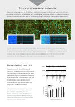
Dissociated neuronal networks Neuronal cultures grown on HD-MEA are used to investigate fundamental properties of brain processing, to study the physiological and pathological functional activity of cultured models on primary or derived cell-lines and for developing drug-screening or toxicological applications. Activity maps and representative signal shapes (bars: 100 ms, 100 µV) of network events occurring in two different dissociated culture models. Left: hippocampal neurons from P0 rats at 14 DIVs (courtesy of Ms. Sinem M. Sertel, University Medical Center, Göttingen). Right: embryonic cortex...
Open the catalog to page 6
Track spontaneous and electrically evoked activity patterns Investigate network activity and connectivity at a micrometre spatial scale with millisecond time resolution, which resembles a standard imaging technique, but is completely label-free. spontaneous bursting activity stimulus-evoked activity Two examples of spatio-temporal propagating patterns in hippocampal cultures. Top: spontaneous synchronous bursting activity. Bottom: evoked response to a biphasic electrical stimulus delivered to the bottom-left corner of the array (courtesy of L. Berdondini NetS3Lab, Fondazione Istituto Italiano...
Open the catalog to page 7
Brain slices BioCAM X and its HD-MEAs with 4096 electrodes allow the researcher to visualise both spiking activity and field potential propagation over large brain circuits (up to 26mm2). Activity map from the 64 by 64 electrode array and examples of the quality of the signals (bars: 100 ms, 100 µV) acquired from a rat cortico-hippocampal slice (left) and a mouse cerebellum tissue (right). Large brain tissues under control Monitor spontaneous/evoked activity patterns propagating over different brain regions. Superimposition of a chemically induced inter-ictal event on the cortico-hippocampal...
Open the catalog to page 8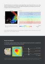
BioCAM X enables the simultaneous monitoring of large areas of neuronal tissue over a long period, thus allowing you to explore the spatial heterogeneity and temporal synchronicity of signals within connected brain areas. CA1 gcl: granule cell layer; slm: stratum lacunosum moleculare; sr: stratum radiatum; sp: stratum pyramidale; so: stratum oriens. Spatial distribution (right) and temporal occurrences (left) of fast ripples detected in the hippocampus and DG (adapted from F. Ortiz, R. Gutiérrez, SfN 2016). Focus on details The HD-MEA’s spatial resolution finely resolves signals coming from dendritic...
Open the catalog to page 9
Retina Either spontaneous or light-induced activity from the explanted retinas of different animals (e.g. murine, salamander, primates, etc.) can be recorded with the BioCAM X HD-MEA system. Mouse retina displaced on the HD-MEA. Colour map activity shows ganglion cell activation (on the right signal amplitude examples; bars: 100 ms, 500 µV) and axonal propagation toward the optic disk (courtesy of E. Sernagor and G. Hilgen, The Institute of Neuroscience, Newcastle, UK). Pan-retinal recording The spatial extent (from 7.1 to 26.2 mm2) of HD-MEAs allows long range interactions and heterogeneous...
Open the catalog to page 10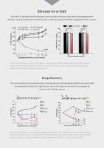
BioCAM X is the ideal tool to develop in vitro models of severe long-term neurodegenerative diseases, such as Alzheimer’s and Parkinson’s, with increased sensitivity compared to other assays. Time of exposure (hours) Sensitivity of HD-MEA to low dose Ap-oligomer concentration (100 nM). While the HD-MEAs show strongly impaired functional activity (left, red curve), the MTT assay does not show significant cell death (right) (adapted from Amin et al., Scientific Reports 2017). Drug discovery The rescue effects of neuroprotective compounds can be evaluated in label-free assays with unprecedented...
Open the catalog to page 11