- Catalogs
- AB-CT - Advanced Breast-CT
- DIEurope Feb/Mar 20: Spiral Breast CT: an innovative technology for high resolution real 3D breast imaging without compression
- Company
- Products
- Catalogs
- News & Trends
- Exhibitions
DIEurope Feb/Mar 20: Spiral Breast CT: an innovative technology for high resolution real 3D breast imaging without compression
1 /3Pages

DIEurope Feb/Mar 20: Spiral Breast CT: an innovative technology for high resolution real 3D breast imaging without compression
1 /3Pages
Catalog excerpts
Breast imaging Spiral Breast CT: an innovative technology for high resolution real 3D breast imaging without compression By E Bärnklau-Gooriah, V Ruth, Dr. C Steiding & Dr. D Kolditz. Even when using images acquired by the latest high-tech breast imaging devices and read by experienced radiologists, the accurate diagnosis of early breast cancer remains challenging. The well-established conventional diagnostic breast imaging modalities all have well-known limitations. In contrast, 3D imaging of the breast at high isotropic resolution offers clear advantages. A dedicated breast CT system, nu:view developed by the German company AB-CT – Advanced Breast-CT, incorporates innovative technology that has the potential to take breast imaging to a new level. The new system provides high resolution 3D imaging of the breast at a very low radiation dose and without the need for any compression. To achieve this, nu:view uses state-of-the-art direct conversion photon-counting detector technology combined with a spiral CT acquisition concept. The CE-marked scanner is commercially available and is already in use with patients in Europe. This article gives an overview of the breakthrough technology as well as the clinical experience already obtained with the system in the University Hospital of Zurich, Switzerland. SPIRAL CT WITH PHOTONCOUNTING DETECTOR The design of the new nu:view scanner allows compression-free imaging of one breast at a time. To do this, the breast CT system uses a rotating gantry on which the X-ray tube and detector are mounted. During the image acquisition process, the gantry rotates around the breast in a downwards-oriented spiral trajectory. In the course of a single 360° rotation of the gantry around the breast, up to 2,000 projection images are acquired. A full spiral scan takes as little as 7 The Authors Eva Bärnklau-Gooriah, Veikko Ruth, Christian Steiding PhD, Daniel Kolditz PhD AB-CT - Advanced Breast-CT GmbH Erlangen, Germany email: [email protected] – 12 seconds. Exceptional image quality at a low radiation dose is possible, because the data acquisition is carried out by a dedicated, curved single photon-counting detector, which is the result of AB-CT’s long-term technical collaboration with the Swedish company Direct Conversion. The nu:view scanner is the world’s first CT in clinical use equipped with a direct converting detector. It features a pixel size of (100 µm)² and a frame rate of up to 1 000 Hz. Unlike traditional scintillation methods used in conventional CTs which convert X-rays into scattered light, in the nu:view photon detector, every single photon is directly converted into electrical energy, thus avoiding scattering effects and resulting in a much higher image quality. The unique combination of the detector’s D I highly sensitive cadmium telluride (CdTe) material with its dedicated shape allows for highest image quality and dose efficiency. High patient comfort The scanner is only 1.10m high which is easily accessible by the patient with the help of a step. The patient table is protected with lead foil sheeting, thus preventing any unintended radiation reaching the patient. For the examination procedure, the patient positions herself prone on the scanner table with the breast to be examined conveniently placed into the aperture. This non-compressive approach keeps the female breast in its natural shape – not only does this avoid the problem of superimposed tissue, but it also makes the examination procedure totally pain-free for the patients. Consequently the patients feel more reassured in a situation which, inevitably, is often highly stressful. A small supporting pillow and footrest may be used to help stabilize the position of the patient who can breath normally during image acquisition. The entire examination process only takes between 3 – 5 minutes, from patient positioning on the scanner table; double-checking of the breast positioning by the radiographer; selection and confirmation of the scan parameters; the scan itself; right through to the final dismounting of the patient from the scanner table. The overall examination time may be slightly extended if contrast media is administered. The actual dataacquiring scan itself takes only 7– 12 seconds, depending on the length of the breast. Scan parameters are adjustable to accommodate various clinical requirements and patient typ
Open the catalog to page 1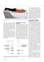
nu:view offers two out-of-the-box protocols for acquisition and reconstruction: a Standard mode with a voxel size of (300 µm)³ and a HighRes mode with (150 µm)³ voxel size. Although the choice of these protocols always depends on individual circumstances, with the ultimate decision being made by the radiologist, the Standard mode is typically the preferred method for scrolling through soft tissue, before switching to HighRes mode e.g. for a more granular analysis of microcalcifications. The nu:view dedicated breast CT scanner is highly appreciated by the patients because no breast compression...
Open the catalog to page 2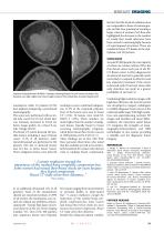
Breast imaging the fact that the levels of radiation dose are comparable to those of mammography and the clear potential of reaching a larger cohort of women, Prof. Boss also highlighted the decrease in the number of recalls that would otherwise have been caused in mammography because of superimposed structures, These are avoided in breast CT, thanks to the multiplanar, real 3D pictures. Conclusion Suspicious microcalcifications BI-RADS 4. Histology: sclerosing adenosis. Top Left: coronal view. Bottom Left: transverse view Right: sagittal view. Clinical images courtesy of Prof. Andreas Boss,...
Open the catalog to page 3All AB-CT - Advanced Breast-CT catalogs and technical brochures
 Flyer nu:view
8 Pages
 OnePager Overview
1 Page
 Showroom
1 Page
 nu:view References
19 Pages
 Testimonials from patients
2 Pages
 DIEurope Breast CT Leiden
4 Pages


Archived catalogs
 nu:view
8 Pages