- Catalogs
- AB-CT - Advanced Breast-CT
- DIEurope Jun 21: Breast CT in women with breast implants
- Company
- Products
- Catalogs
- News & Trends
- Exhibitions
DIEurope Jun 21: Breast CT in women with breast implants
1 /3Pages

DIEurope Jun 21: Breast CT in women with breast implants
1 /3Pages
Catalog excerpts
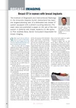
BREAST IMAGING Breast CT in women with breast implants The Institute of Diagnostic and Interventional Radiology in the University Hospital Zurich, Switzerland has been the longest-standing user of a dedicated new breast CT system equipped with a photon-counting detector. The team have just published a paper on the use of the new system in patients with breast implants [1]. We spoke to Prof. Andreas Boss, Senior Consultant responsible for breast imaging. Before we get to the use of the new breast CT system in detail, please describe the breast imaging unit at the University Hospital Zurich and the Swiss breast screening system in general. OK. Let’s start with our imaging equipment: this includes a mammography/tomosynthesis unit from Siemens Healthineers; a handheld US device and an automated 3D breast ultrasound (ABUS) both from GE; and two 3T MRI from Siemens Healthineers. In addition we have an AI-derived software system from the company b-rayZ for the analysis of breast density and mammography image quality. And as you mentioned we also have the new spiral breast-CT system from AB-CT. All this equipment is put to a lot of use, since we see a lot of patients. Each year we carry out 2,500-3,000 mammographies and approximately 700 breastCT examinations. In Switzerland, the practical implementation of organized QC mammography breast cancer screening programs is the responsibility of the canton. Overall, approximately 60% of Swiss women in the age range 50-70 have access to such screening programs. However, many cantons — particularly in the German speaking part of Switzerland, including the canton of Zurich — have not yet implemented systematic organized screening programs. In these areas women who want to be screened typically have to resort to “opportunistic” screening examinations in a radiology institution (which is not necessarily covered by public health insurance). Depending on the particular risk profile 16 and the risk awareness of the individual patient, opportunistic mammography breast cancer screening starts at the age of 40 years, typically with examinations every two years. One advantage of opportunistic breast cancer screening mammography compared to the organized screening program is that additional breast ultrasound can be carried out in patients with dense breasts. It is well known that the sensitivity of conventional mammography drops substantially in patients with dense breasts. In such cases adjunct ultrasound examinations significantly increase the detection rate for breast cancer. In our institution, a “lean” workflow is implemented. Thus, immediately after the screening mammography examination, the images are analyzed with the “b-box” AI medical device from the Swiss company b-rayZ to assess the breast density according to the ACR BI-RADS system. For women with the highest breast density categories, i.e. categories c-d, an additional ABUS examination is carried out by the technician. We hear a lot about AI in radiology these days but an excellent example of its usefulness is in the determination of mammographic breast density. The BI-RADS breast density system doesn’t use a quantitative score scale but instead uses both the amount and the distribution of breast tissue to attribute density to one of four categories (a, b, c, d). The result is that software tools relying on quantitative measurements of the amount of breast density are miscalibrated to a ACR BI-RADS score. Artificial Intelligence algorithms such as D I Prof Andreas Boss is Senior Consultant responsible for breast imaging at the Institute of Diagnostic and Inter-ventional Radiology. [email protected] those in the b-box system can significantly improve classification, Women at highest risk, e.g. with known BRCA mutations, are recommended to follow a more intense screening schedule, according to the guidelines of the Swiss Cancer League, which suggest that such women should undergo yearly breast MRI examinations. All the above is a simplified description of our standard screening procedures, but a problem is that a large number of women in Switzerland are unwilling to undergo the often painful breast compression required for mammography or tomosynthesis. For such patients, we now offer breast cancer screening examination using the nu:view breastCT system from AB-CT. [Figure 1] Figure 1. The nu:view Mamma-CT system was developed and is produced by the German company AB-CT - Advanced Breast CT. The design of the new scanner allows compression-free imaging of one breast at a time. To do this, the breast CT system uses a rotating gantry on which the X-ray tube and photon-counting detector are mounted. During the image acquisition process, the gantry rotates around the breast in a downwards-oriented spiral trajectory. In the course of a single scan up to 12,000 projection images are acquired. A full spiral scan takes as little as 7 – 12 seconds. The radiation dose is similar to that of conventional mamm
Open the catalog to page 1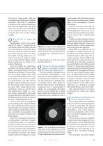
The breast-CT image quality is high with microcalcifications being clearly visualized. In addition, if the breast-CT examination is carried out with contrast enhancement, both soft tissue enhancement of the breast cancer and associated microcalcifications can be visualized at the same time, which cannot be done with any other imaging modality. Now let’s turn to women with implants. Approximately 10-20% of the patients referred for breast-CT imaging have silicone implants, either for cosmetic purposes or for reconstruction after breast cancer treatment. Overall the number of women with silicone...
Open the catalog to page 2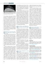
BREAST IMAGING Figure 4. Single microcalcification seen in the axial plane in a 65-yr old woman with bilateral gel implants. Image adapted with permission from Ref 1. to the reading of the datasets. Initially, detecting microcalcifications in the 3D datasets was a challenge, as they appear less prominent than in mammography because of the high isotropic spatial resolution. In practice we adapted to this by creating maximum-intensity-projections with our PACS viewing system with slice thicknesses of the order of 2-3 mm, which allows microcalcifications to be detected with an accuracy similar to...
Open the catalog to page 3All AB-CT - Advanced Breast-CT catalogs and technical brochures
 Flyer nu:view
8 Pages
 OnePager Overview
1 Page
 Showroom
1 Page
 nu:view References
19 Pages
 Testimonials from patients
2 Pages
 DIEurope Breast CT Leiden
4 Pages


Archived catalogs
 nu:view
8 Pages