- Catalogs
- AB-CT - Advanced Breast-CT
- Spiral Breast CT: Going Further with Photon Counting (Source: DIEurope)
- Company
- Products
- Catalogs
- News & Trends
- Exhibitions
Spiral Breast CT: Going Further with Photon Counting (Source: DIEurope)
1 /4Pages

Spiral Breast CT: Going Further with Photon Counting (Source: DIEurope)
1 /4Pages
Catalog excerpts
Spiral Breast CT: Going Further with Photon Counting In this article we introduce the innovative technical features of the nu:view breast CT system and describe recent clinical findings and the likely indications of this new equipment. Offering significant advantages over other breast imaging modalities thanks to its innovative design and performance characteristics, dedi ca ed breast CT is so far the only 3D t breast imaging method that has been shown to reliably detect microcalcifications and mass lesions without any risk of obscuring lesions because of superimposed tissue. Contrast-enhanced imaging with the new technique enables sensitive detection of malignant mass lesions as well as of non-mass lesions. Overall, dedicated breast CT has been shown to be an appropriate modality for a number of clinical indications. Problem-solving is one such application; lesions that are ill-defined by other imaging modalities can easily be identified by 3D spiral breast CT and additional tumor characteristics assessed through contrast enhancement. Another field of application is preoperative staging of established breast cancer cases in order to determine the optimal therapeutic strategy. However, probably the largest potential of dedicated breast CT lies in screening, where currently many women drop out of standard screening mammography programs because of the discomfort and pain associated with the breast compression involved in conventional mammography. This problem is absent in spiral breast CT, which does not involve breast compression. Breast imaging is the unavoidable backbone technology in the diagnosis of breast cancer, with the gold standard imaging method currently being digital mammography. Several other imaging modalities such as ultrasound and breast MRI also have well-established roles and recent methods such as breast tomosynthesis are increasingly being used in breast imaging. In addition, the potential of newer imaging modalities such as contrast-enhanced spectral mammography (CESM) or breast CT is under active evaluation to determine their optimal role in the workup of breast lesions. Recently a dedicated breast CT system, nu:view, developed by the German company AB-CT (Advanced Breast CT) has become commercially available in Europe, after obtaining CE certification in 2018. A prototype of the scanner was installed as early as 2019 in our breast radiology department at Erlangen University Hospital for preclinical studies and since then it has been also used for routine clinical examinations. So far we have carried out more than 200 clinical examinations with the new device, principally for advanced diagnostic purposes. Current breast cancer diagnostics The field of breast diagnostics is already supplied with many different imaging techniques, so on the face of it, a legitimate initial question is to wonder whether yet anoth- The Authors Dr. Matthias Wetzl · Dr. Sabine Ohlmeyer · Dr. Evelyn Wenkel University Hospital Erlangen · Department of Radiology · Maximiliansplatz 3 · 91054 Erlangen · Germany www.uk-erlangen.de Corresponding author Dr. Sabine Ohlmeyer · [email protected]
Open the catalog to page 1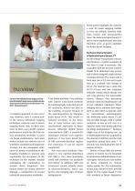
Image: Universitätsklinikum Erlangen These points highlight the need for a new 3D breast imaging modality that can reliably visualize both mass lesions and microcalcifications. The dedicated spiral breast CT meets these requirements and may therefore serve as good candidate for future breast imaging. Technical characteristics of dedicated spiral breast CT The team of the multimodal breast imaging unit of the University Hospital Erlangen have accumulated extensive clinical experience with the new nu:view scanner from AB-CT – Advanced Breast-CT. er imaging approach is even necessary. However, even...
Open the catalog to page 2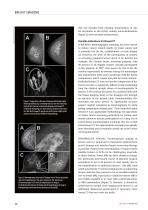
Image: Universitätsklinikum Erlangen that can visualize both imaging characteristics of ductal carcinoma in situ (DCIS), namely microcalcifications [Figure 2], and non-mass enhancement. Possible indications for Breast CT Figure 1. Images from a 59-year old woman with bloody nipple discharge without any correlating lesion in two-view mammography (A, B). Contrast-enhanced dedicated spiral breast CT revealed an enhancing non-mass lesion in the lower outer quadrant (circled in image C), which was confirmed as a high-grade ductal carcinoma in situ (DCIS) on histopathology. Figure 2. Mammography and...
Open the catalog to page 3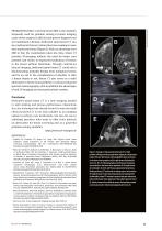
Image: Universitätsklinikum Erlangen PROBLEM-SOLVING: Currently breast MRI is the modality frequently used for problem solving in breast imaging cases where analysis is difficult and precise diagnosis not yet established. Likewise, dedicated spiral breast CT can be a method of choice in clinical decision-making in cases with equivocal lesions [Figure 3]. With an advantage over MRI in that the examination takes less time, breast CT provides 3D-imaging without the need for breast compression and results in improved localization of lesions in the breast without distortions. Through contrast-enhanced...
Open the catalog to page 4All AB-CT - Advanced Breast-CT catalogs and technical brochures
 Flyer nu:view
8 Pages
 OnePager Overview
1 Page
 Showroom
1 Page
 nu:view References
19 Pages
 Testimonials from patients
2 Pages
 DIEurope Breast CT Leiden
4 Pages


Archived catalogs
 nu:view
8 Pages