- Catalogs
- Akoya Biosciences
- PhenoImager HT Automated Quantitative Pathology Imaging System
- Products
- Catalogs
- News & Trends
- Exhibitions
PhenoImager HT Automated Quantitative Pathology Imaging System
1 /6Pages

PhenoImager HT Automated Quantitative Pathology Imaging System
1 /6Pages
Catalog excerpts

PRODUCT NOTE | PhenoImager HT Automated Quantitative Pathology Imaging System AKOYA BIOSCIENCES' We've rebranded some of our products. Phenoptics™ is now PhenoImager™ and Vectra® Polaris™ is now PhenoImager™HT. THE SPATIAL BIOLOGY COMPANY* PhenoImager HT Automated Quantitative Pathology Imaging System Quantitative Pathology Imaging and Analysis KEY FEATURES: • State of the art multispectral imaging with spectral unmixing to easily detect and measure multiple overlapping biomarkers within a single tissue section • High speed digital whole-slide scanning at 10X - 40X in brightfield or fluorescence • Enclosed system with built-in touchless automation allows users to safely visualize, analyze, quantify, and phenotype immune cells in situ with maximum reliability • Trainable machine-learning algorithms using inForm® software allowing for automatic identification of specific cell and tissue types MORE ACCURATELY QUANTIFY CELLULAR INTERACTIONS IN TISSUE SAMPLES The PhenoImager HT™ (formerly Vectra® Polaris™) Automated Quantitative Pathology Imaging System is a new class of tissue imager which provides unparalleled speed, performance, and versatility for visualizing, analyzing, quantifying, and phenotyping immune cells in situ in Formalin Fixed Paraffin Embedded (FFPE) tissue sections and TMAs to advance disease research. PhenoImager goes beyond basic functions, like whole-slide, brightfield (BF), and fluorescence (FL) imaging. It integrates the power of multispectral imaging (MSI) and spectral unmixing in a simplified workflow, and is part of our PhenoImager HT translational solution. With our patented and proven MSI technology with spectral unmixing, you can capture the multiple interactions occurring between cells without the interference of autofluorescence as the signals are unmixed from one another. That means you can have confidence in accurately quantifying the interactions that are really occurring in the biology. Multispectral imaging on the PhenoImager HT can now be applied across a whole slide for up to 7 colors and unmixed in less than 8 minutes with our state-of-the-art onboard spectral unmixing technology, enabling the biology to be explored at multiple scales, from cell-to-cell interactions to the macroscopic tissue architecture. The whole slide multispectral imaging capability creates a simpler and more robust workflow as fields of view do not need to be selected so there's no selection bias and you have a whole slide record so that you can easily re-analyze imagery as new understanding emerges. With the PhenoImager HT, you can also generate unmixed annotated regions of interests of up to 9 colors using our renowned liquid crystal tunable filter (LCTF) technology. Combined with a fit-for-need selection of software analysis options and PhenoCode Signature staining panels, you can identify and quantify multiple targets and reveal spatial relationships within a digital workflow for faster, easier and more informative studies. UNDERSTAND BIOLOGICAL INTERACTIONS ACROSS MANY APPLICATIONS The PhenoImager HT Automated Quantitative Pathology Imaging System is a cutting-edge tool for revealing the cell-to-cell biology driving tumor progression, immune interactions and response to therapeutic interventions. By combining tissue imaging with multiplexing, the PhenoImager HT system can capture this biology with a workflow that supports the volume and high- throughput demands of clinical research studies.
Open the catalog to page 1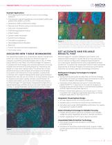
PRODUCT NOTE | PhenoImager HT Automated Quantitative Pathology Imaging System Example Applications • Phenotyping immune cells for cancer immunology research • Transduction signaling pathway activity (pAKT, pERK, pS6, p13K/mTOR, MAPK, or EGFR) • Apoptosis and/or proliferation assays • Necrosis and fibrosis using conventional stains • Cell cycle characterization • DNA damage determination • Inflammation • Lymph node metastasis • Autoimmune diseases • Transplant acceptance • Neurodegenerative diseases • RNA-ISH • Tumor microenvironment exploration • And many more! DI S COV ER N EW T ISSUE BIOMARKERS...
Open the catalog to page 2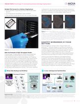
AKOYA BIOSCIENCES* PRODUCT NOTE | PhenoImager HT Automated Quantitative Pathology Imaging System Multiple File Formats for a Variety of Applications • 16-bit extended range data format to reduce saturation rescans • Unmixed data for simplified 4+ plex workflow • 8 bit standard range data format with compact file sizes Meet the Demands of High-Throughput Studies PhenoImager HT goes beyond conventional brightfield and fluorescence whole-slide scanning. Utilizing the most advanced optics, mechanical design, and imaging sensors; PhenoImager HT delivers both high performance and high-throughput for...
Open the catalog to page 3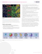
PRODUCT NOTE | PhenoImager HT Automated Quantitative Pathology Imaging System • User-trained feature recognition algorithms allow automatic identification of specific tissue types based on tissue morphology • Adaptive Cell Segmentation reliably identifies individual cell types in densely packed, complex morphologies regardless of staining heterogeneity and background levels for accurate phenotyping and downstream spatial analysis in phenoptr • Allows quantitative per-cell analysis of biomarker expression in tissue sections and TMAs • Automatically classifies cell phenotypes using machinelearning...
Open the catalog to page 4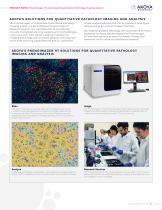
PRODUCT NOTE | PhenoImager HT Automated Quantitative Pathology Imaging System A KOYA’S SO LUT IONS FOR QUANT I TAT I V E PAT HOLOGY I MA G I NG A ND A NALYS I S The PhenoImager HT Automated Quantitative Pathology Imaging System is a part of Akoya’s PhenoImager HT Research Solution. Our complete end-to-end solution includes multiplexed staining reagents and methodologies, instrumentation with industry leading multispectral imaging technology with on-board spectral unmixing and whole slide scanning capabilities (BF and FL), advanced image analysis software that can be trained to phenotype cells...
Open the catalog to page 5All Akoya Biosciences catalogs and technical brochures
 Opal Storage and Stability
2 Pages
 Anti-Hu Ki67
2 Pages
 TSA® Research Reagents
12 Pages
