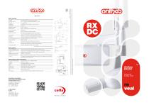
RX DC Wired | Anthos
1 /2Pages

RX DC Wired | Anthos
1 /2Pages
Catalog excerpts
Anode current Voltage at X-ray tube Exposure times Source-skin distance Irradiated field Ø 60 mm and Ø 55 mm (with round cone) Additional collimators 35 x 45 mm (with rectangular cone for size 2 sensors) 31 x 41 mm and 22 x 35 mm, for size 1 and size 0 sensors Power supply Duty Cycle Continuous operation with self-adjustment up to 1s/80s total Total filtration Serial with USB adapter available in various lengths (*) values depend on the country where the product is marketed. 400/ 600/ 900 (15.7/ 23.6/ 35.4) iRYS (compliant with ISDP©10003:2020 in accordance with EN ISO/IEC17065:2012 certificate number 2019003109-3) and iPad iRYS viewer App (free) Protocols supported in iRYS DICOM 3.0, TWAIN, VDDS DICOM Node Connectivity iRYS - IHE compliant (Print; Storage Commitment, SR document; WorkList; MPPS; Query/Retrieve) iRYS feature to associate exposure parameters with the X-ray images of each examination (exportable in PDF or CSV format) Use a power adapter of a power suitable for the video card in use Power supply Display settings Discrete 3D Video Card or integrated GPU Graphics card Intel Core i3 or higher Hard Disk Microsoft® Windows® 10, 11 Professional 64 bit RX DC minimum system requirements Supported operating systems iCapture for automatic saving of RX DC exposure parameters on PC Image management software (for PC) Viewing on a handheld device with digital archive option on PC via iRYS software which can be automated via the “RX DC connect” (optional) accessory Dose delivered Standard (wall mounted) or Mobile (on portable cart) Arms (for Standard version only) Available in 3 lengths: 40 cm – 60 cm – 90 cm Focal spot Constant-potential, microprocessor-controlled Working frequency BU MEDICAL EQUIPMENT SEDE LEGALE ED AMMINISTRATIVA HEADQUARTERS Cefla s.c. Via Selice Provinciale, 23/a 40026 Imola - BO (Italy) tel. +39 0542 653111 fax +39 0542 653344 STABILIMENTO PLANT Via Bicocca, 14/c 40026 Imola - BO (Italy) tel. +39 0542 653441 fax +39 0542 653601 The images and technical specifications shown in this catalog are for indicative purposes only. As part of ongoing technological updates, technical specifications may be subject to changes without prior notice. In accordance with current regulations, in non-EU areas some products, as well as certain technical specifications, may have different availability and configurations. We encourage you to always contact your local distributor for up-to-date technical specifications, availability and configurations.
Open the catalog to page 1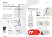
X-ray imaging that is precise, practical and versatile We’ve designed and built the instruments your surgery’s been waiting for: practical, high definition, ergonomic and versatile. Instruments that make work easier and more professional, that improve dentist-patient relations RX DC X-RAY IMAGING UNIT thanks to immediate diagnosis and real-time high definition imaging. Solutions that adapt to the dentist’s work, boosting the surgery’s diagnostic capabilities and improving the quality of the work provided. Higher performance and maximum ergonomics. Thanks to the protractor with graduated scale,...
Open the catalog to page 2All Anthos catalogs and technical brochures
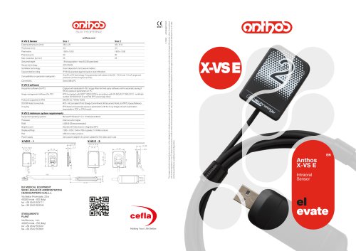 X-VS E | Anthos
2 Pages
 A | Anthos
6 Pages
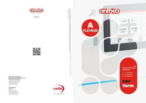 A Platinum | Anthos
6 Pages
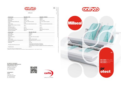 Millseal | Anthos
2 Pages
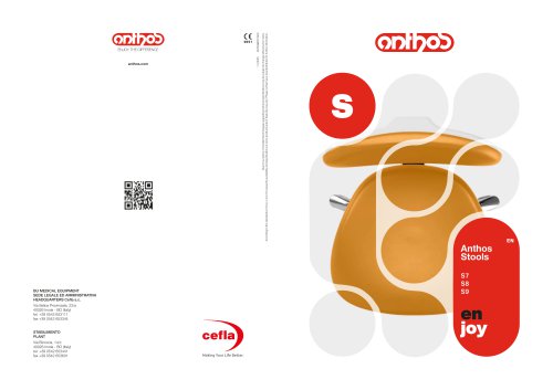 Stools | Anthos
6 Pages
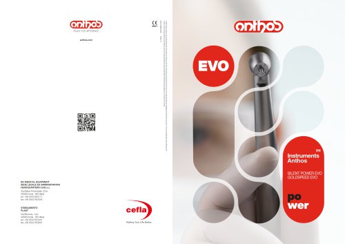 Instruments | Anthos
4 Pages
 Cart | Anthos
8 Pages
 R7 | Anthos
24 Pages
 MTC View | Anthos
4 Pages
 L6 - L9 | Anthos
26 Pages
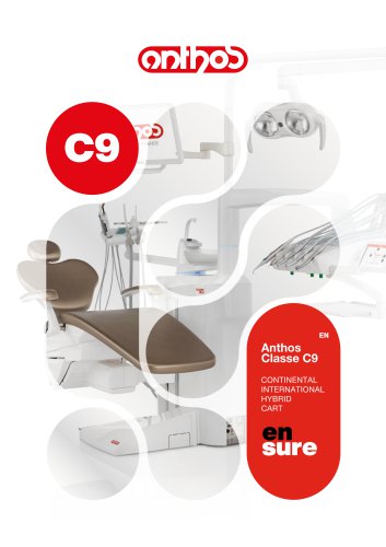 C9 | Anthos
23 Pages
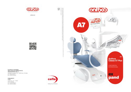 A7 | Anthos
20 Pages
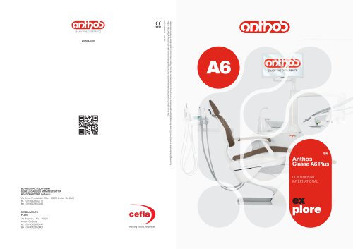 A6 | Anthos
18 Pages
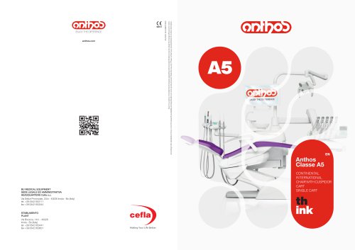 A5 | Anthos
16 Pages
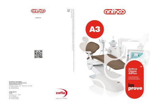 A3 | Anthos
12 Pages
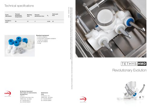 Tethys HMD | Anthos
2 Pages
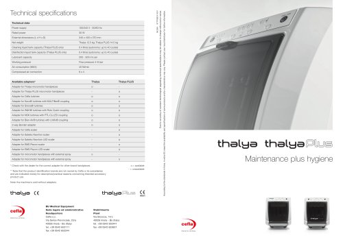
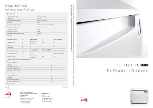 Tethys H10 Plus | Anthos
6 Pages
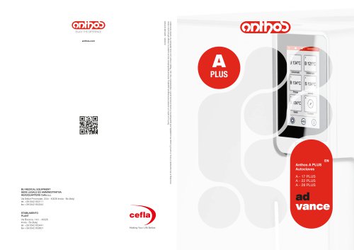 A Plus | Anthos
6 Pages
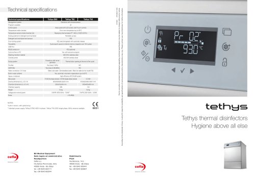 Tethys | Anthos
4 Pages
- Sterilizer
- Surgical lighting system
- Steam sterilizer
- LED surgery light
- Analysis software
- Syringe
- Ceiling-mounted surgical light
- Benchtop sterilizer
- Automatic sterilizer
- Stainless steel sterilizer
- Dental treatment unit
- Visualization software
- Radiology software
- Flat panel detector
- Control software
- Stool with backrest
- Dental unit with light
- Windows software
- Drill
- Micromotor