


Foot and Ankle

Foot and Ankle
The document provides a detailed guide on the Osteochondral Autograft Transfer System (OATS) for treating osteochondral lesions in the foot and ankle, focusing on the talus and metatarsal regions. It covers clinical outcomes, surgical techniques, and post-operative protocols.
- Osteochondral Autograft Transplantation for Type-V Cystic Osteochondral Lesions of the Talus: A study of 50 patients showed a 90% success rate with good-to-excellent outcomes using grafts from the ipsilateral knee.
- Bone-Cartilage Transplantation from the Ipsilateral Knee: A prospective analysis of 43 patients demonstrated successful integration of grafts with surrounding cartilage, indicating promising results for treating local cartilage lesions.
- Osteoarticular Transplantation of Lesser Metatarsal Articular Deficits: A preliminary study showed favorable outcomes with significant improvements in AOFAS scores post-surgery.
- Osteochondral Autograft Transfer of the First Metatarsal Head: A case report highlighted successful graft integration and functional joint range of motion at 12 months post-operation.
- Osteochondral Autografting for Cystic Talar Lesions: The procedure involves determining lesion size, choosing the appropriate approach (medial or lateral), and using specific tools for graft harvesting and insertion.
- Metatarsal Technique: Involves a detailed process for addressing defects in the metatarsal head, including incision, defect measurement, and graft insertion.
Aftercare includes prophylactic antibiotics, pain management, and a structured weight-bearing schedule over nine weeks, followed by physical therapy and rehabilitation.
- Proper lesion visualization and access are crucial, sometimes requiring a malleolar osteotomy.
- Accurate measurement and alignment during graft insertion are essential for successful outcomes.
- Post-operative care is critical to ensure graft integration and patient recovery.
Layered closure is performed with careful attention to the capsular closure of the joint to ensure proper reapproximation. For operations on a lesser metatarsophalangeal joint, the toe is splinted in neutral or slight plantar flexion to facilitate capsule healing.
Initially, the patient is placed in a posterior splint and instructed to remain nonweight-bearing, returning in two weeks for suture removal. A nonweight-bearing below-the-knee cast is applied at the first postoperative visit, with a follow-up in three weeks. At five weeks postoperatively, a radiograph checks the graft's healing progress, and the patient is either placed in a cast brace or a below-the-knee cast, remaining nonweight-bearing for another three weeks. At eight weeks, another x-ray is taken. If healing is confirmed, the patient begins partial weight-bearing for three weeks, followed by two weeks of full weight-bearing in a cast brace, eventually progressing to shoe gear as tolerated. High-impact activities are only permitted after five months of recovery.
Disposables include Small Joint OATS Sets in various sizes (6 mm, 8 mm, 10 mm) and Talus Allograft OATS Set. Each set includes necessary instruments such as a Graft Delivery Tube, Graft Driver, Tamp, and others. The document lists U.S. Patent numbers and emphasizes that the technique description is an educational tool for licensed medical professionals, who should use their judgment and experience in product usage.
Catalog excerpts
OATS® for the Foot & Ankle Surgical Technique OATS for the Foot & Ankle
Open the catalog to page 1
OATS for the Foot & Ankle Scientific Support Outcome of Osteochondral Autograft Transplantation for Type-V Cystic Osteochondral Lesions of the Talus P. E. Scranton, Jr., M.D.; C. C. Frey, M.D.; and K.S. Feder, M.D. Journal of Bone and Joint Surgery. British Volume, Vol 88-B (Issue 5) 2006;614 19. –6 “The treatment of osteochondral lesions of the talus has evolved with the development of improved imaging and arthroscopic techniques. However, the outcome of treatment for large cystic type-V lesions is poor, using conventional grafting, debridement or microfracture techniques. This retrospective...
Open the catalog to page 3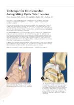
Technique for Osteochondral Autografting Cystic Talar Lesions Pierce Scranton, M.D., Seattle, WA; and Mark Easley, M.D., Durham, NC The patient is supine and the appropriate limb is prepped and draped with a high thigh tourniquet. An arthroscopic leg holder is not necessary. A general or regional (spinal) anesthetic is required. The lesion size and OATS drill size can be determined from direct measurement or from CT or MRI (these should be used for planning only, with the kit not opened, until the defect size is clinically confirmed). The harvester in the kit will match the drill size exactly....
Open the catalog to page 4
The Drill Tip Guide Pin is then overdrilled with the appropriate size Cannulated Headed Reamer to a depth of at least 12 mm. Note: A second or even third core can be punched adjacent to the first hole with the recipient corer in large lesions where “nesting” of grafts is required. The cannulated OATS Alignment Rod is introduced over the Drill Tip Guide Pin for a depth measurement. This should be tapped to the base of the drill hole for an accurate depth measurement. Knowing the diameter and depth of the talar hole, the wound is covered with a saline-dampened sponge and attention is directed to...
Open the catalog to page 5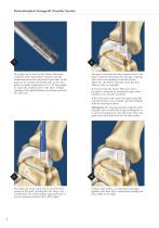
Osteochondral Autograft Transfer System The graft can be sized in the Donor Harvester using the graft visualization windows and the measurements on the end of the metal tube, or the graft can be extruded and sized with a ruler. The graft is carefully rongeured to 11.5 2 mm length –1 to match the recipient hole in the talus. A slight tapering of the graft facilitates its introduction into the talar hole. The graft is inserted into the recipient hole in the talus in optimal orientation for articular congruity. This can be accomplished with the Donor Harvester, the Donor Harvester with the clear...
Open the catalog to page 6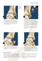
Surgical Technique Malleolar Osteotomy (if required for access) The medial malleolus is predrilled with two 0.045” pins at slightly divergent angles to help prevent proximal slippage of the medial malleolus during screw insertion. These pins are overdrilled with the 3.4 mm cannulated Trim-It™ Drill Bit across the medial malleolus and into the tibial plafond. The holes are then tapped. Using intraoperative fluoroscopy or by direct vision, create a 45° osteotomy cut from the superior medial malleolus down to the junction of the tibial plafond and medial colliculus leaving the last 1/8 of the bone...
Open the catalog to page 7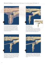
Metatarsal Technique (Matthew Rockett, DPM, Houston, TX; and Michael Aquino, DPM, Buffalo, NY) A 4-5 cm incision is created starting just distally to the metatarsophalangeal joint and carried over the joint to a level of the surgical neck of the metatarsal. Subcutaneous dissection is taken down to the level of the periosteum—capsular level. A linear dorsal capsulotomy is performed and the metatarsal head is dissected to expose the metatarsal head and joint. Advance the reamer over the Drill Tip Guide Pin and remove the defect and any related subchondral cystic changes to a minimum depth of 8...
Open the catalog to page 8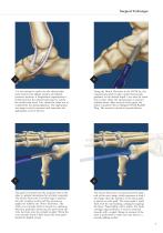
Surgical Technique 3 cm incision is made over the talonavicular –5 joint between the tibialis anterior and tibialis posterior tendons. A longitudinal capsulotomy is performed into the talonavicular joint to expose the medial talar head. Care should be taken not to compromise the spring ligament. The appropriate size tamp is used to measure and determine the appropriate area for harvest. 7 The graft is inserted into the recipient hole in the talus in optimal orientation for articular congruity. The Donor Harvester’s beveled edge is inserted into the recipient socket and firm pressure is applied...
Open the catalog to page 9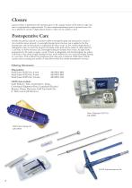
Closure Layered closure is performed with attention given to the capsular closure of the joint to make sure that it is appropriately reapproximated. If a lesser metatarsophalangeal joint is operated upon, the toe is splinted in neutral or slight plantar flexion to allow for the capsule to heal. Postoperative Care Initially, the patient is placed in a posterior splint nonweight-bearing and instructed to return in two weeks for suture removal. A nonweight-bearing below-the-knee cast is applied at the first postoperative visit and the patient is reappointed for three weeks. At five weeks postoperatively,...
Open the catalog to page 10
www.arthrex.com ...up-to-date technology just a click away This description of technique is provided as an educational tool and clinical aid to assist properly licensed medical professionals in the usage of specific Arthrex products. As part of this professional usage, the medical professional must use their professional judgment in making any final determinations in product usage and technique. In doing so, the medical professional should rely on their own training and experience and should conduct a thorough review of pertinent medical literature and the product’s Directions For Use. U.S. PATENT...
Open the catalog to page 12All Arthrex catalogs and technical brochures
 Imaging and Resection Catalog
108 Pages
Archived catalogs
 ACL TightRope® RT - LS0178
2 Pages
 HTO Medial PEEK Implant
6 Pages
 A rthrex Graft Tubes
4 Pages
 Clavicle Plate and Screw System
12 Pages
 For Fracture Treatmen
8 Pages
 Hip Labral Scorpion
1 Page
 Imaging & Resection
80 Pages
 Hand & Wrist
64 Pages
 Shoulder Repair Technology
114 Pages
 Ankle Arthroscopy
11 Pages
 Arthroscopic Hand Instruments
28 Pages
 Foot & Ankle
138 Pages




- Bone plate
- Compression plate
- Planning software
- Suture thread
- Surgery software
- Non-absorbable suture thread
- Arthrex® suture anchor
- Arthrex® non-absorbable suture anchor
- Foot compression plate
- Orthopedic surgery suture thread
- Rotator cuff suture anchor
- Suture button
- Small joint suture anchor
- ACL reconstruction suture button
- Calcaneus compression plate
- Shoulder prosthesis
- Knotless suture anchor
- Preoperative planning software
- Suture needle
- Biceps tendon suture anchor