- Catalogs
- Big Vision Medical
- OCT Opthalmoscope BV1000
OCT Opthalmoscope BV1000
1 /8Pages
OCT Opthalmoscope BV1000
1 /8Pages
Catalog excerpts

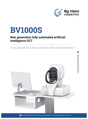
New generation fully automated artificial intelligence OCT A new generation of fully automatic artificial intelligence OCT
Open the catalog to page 1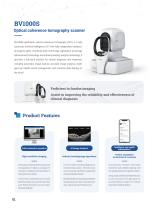
Optical coherence tomography scanner BV1000S ophthalmic optical coherence tomography (OCT) is a fully automatic artificial intelligence OCT with fully independent intellectual property rights. Combined with multi-image registration and image enhancement technology and industry-leading analysis technology, it provides a full-stack solution for clinical diagnosis and treatment, including automatic image capture, accurate image analysis, intelligent eye health record management, and real-time data sharing on the cloud. Proficient in fundus imaging Assist in improving the reliability and effectiveness...
Open the catalog to page 2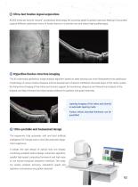
80,000 times per second "second" acceleration technology, the scanning speed is greatly improved, helping to accurately capture different subdivision forms of fundus lesions in a shorter time and obtain high-qualityimages. 0 Hyperfine fundus structure imaging The AI multimodal ophthalmic image analysis algorithm based on deep learning can more finelyobserve the subdivision morphology of various fundus diseases and the development of lesions indifferent structural layers of the retina, realize the hierarchical imaging of the retina and choroid, support the monitoring, diagnosis and hierarchical...
Open the catalog to page 3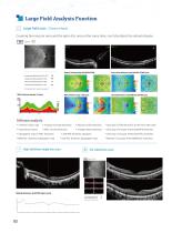
Large field scan (i 2mm x 9mm)Covering the macular area and the optic disc area at the same time, can fully detect the retinal disease. Average choroidal thickness(gm)(73~319) 254 N/A indicates that it could not be analyzed RNFL thickness diameter (3.4mm) Software analysis ♦ Grid map of TSNI thickness of the nerve fiber layer ♦ Grid map of macular 6*6mmILM-RPE thickness ♦ Grid map of macular 6*6mmILM-0PL thickness ♦ Meshes of macular 6*6mmBRM-SCL thickness ♦ 12*9mm fundus map ♦ Average choroidal thickness ♦ Macular foveal thickness♦ Total retinal volume ♦ RNFL circular thickness ♦ Average retinal...
Open the catalog to page 4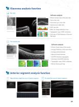
Software analysis ♦ 6*6mm fundus map of the optic disc ♦ Horizontal cup to disc ratio ♦ Vertical cup to plate ratio ♦ RNFL rim sweep thickness curve ♦ Topographic map of RNFL thickness ♦ Grid map of TSNI thickness of the nerv fiber layer ♦ Average thickness of the inner retina ♦ Average retinal thickness ♦ Macular foveal thickness ♦ ILM-RPE thickness topography ♦ Grid map of ILM-RPE thickness ♦ Topographic map of RNFL thickness ♦ Retinal volume grid map
Open the catalog to page 5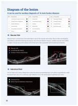
Macular hole is a tissue loss of the entire layer or part of the macular nerve retina. Most of them are idiopathic and related to abnormal vitreomacular traction, and a few are related to trauma. Lamellar macular hole (LMHH) is a layer of tissue loss on the surface of the fovea and is often associated with the epiretinal membrane. On OCT, there was a serous detachment under the retinal neuroepithelial layer, with uniform hyporeflection within the detachment area, and a continuous light reflection zone in the retinal pigment epithelial layer below.
Open the catalog to page 6
(0D) Suspected fundus abnormality, seethe disease section page! (OS) Suspected fundus abnormality, see the disease section page! CAI-OCT was selected into the 8th batch of excellent Domestic medical equipment product list of China Medical Equipment Association CMIAS-3000 was awarded the winning unit of the artificial intelligence medical device Innovation task unveiled by the Ministry of Industry and Information Technology CAI-OCT was awarded the honorary title of "Scientific and Technological Progress in Ophthalmology of China Medical Equipment Association" Cr A cooperative research center for...
Open the catalog to page 7
Technical parameters Chinese NMPA: Jiangxi 20202160485 Note: When selecting the anterior segment mode, the peripheral devices of the anterior segment should be in place; the design and parameters are subject to change without prior notice. Building 6,199 Jinyuan Road, Medpark, 215000 Suzhou, ChinaFollow us on Linkeln: https://www.linkedin.com/company/bigvision-medical/ Website: https://www.bigvisiontech.com/Email: [email protected] (§) +86 400 1 28 0866
Open the catalog to page 8All Big Vision Medical catalogs and technical brochures
 Biometer BVB2000
5 Pages
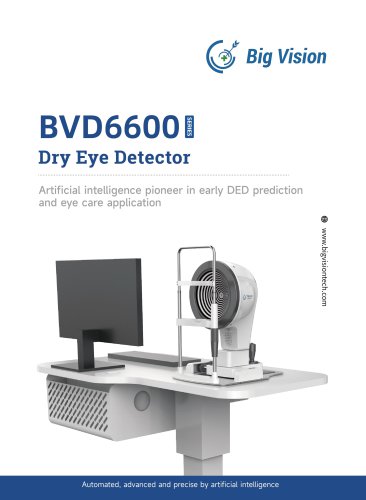 Dry eye BVD6600
8 Pages
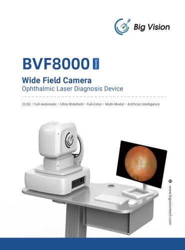 Retinal camera wide field
12 Pages
- Analysis software
- Tablet computer software
- Tablet PC software
- Control software
- Monitoring software
- Diagnostic software
- Fixed ophthalmic examination
- Treatment software
- Data analysis software
- Real-time software
- Ophthalmoscope
- Interpretation software
- Smartphone software
- Screening software
- Retinal camera
- Ophthalmic software
- Ophthalmic biometer
- Eye software
- OCT ophthalmoscope
- Aid software