- Catalogs
- Big Vision Medical
- Retinal camera wide field
Retinal camera wide field
1 /12Pages
Retinal camera wide field
1 /12Pages
Catalog excerpts
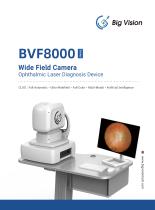
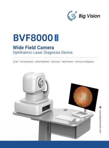
Wide Field CameraOphthalmic Laser Diagnosis Device CLSO : Full-Automatic • Ultra-Widefield • Full-Color • Multi-Modal • Artificial Intelligence
Open the catalog to page 1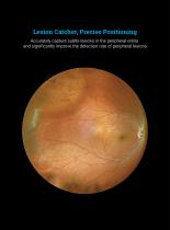
Accurately capture subtle lesions in the peripheral retina and significantly improve the detection rate of peripheral lesions
Open the catalog to page 3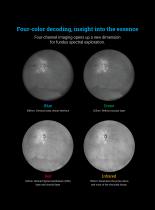
Green 488nm: Vitreous body, retinal interface 525nm: Retinal vascular layer Red Infrared 635nm: Retinal Pigment Epithelium (RPE) 785nm: Penetrates the entire retina layer and choroid layer and most of the choroidal tissue.
Open the catalog to page 4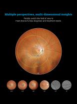
Flexibly switch the field of view to meet diverse fundus diagnosis and treatment needs.
Open the catalog to page 5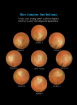
Fundus color photography 9 positions diagram, Construct a panoramic diagnostic perspective.
Open the catalog to page 6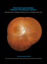
Multi-eye position intelligent stitching to draw a complete fundus map. Three-eye position stitching 260° panoramic stitching, real-time dynamic stitching, automatic stitching, manual stitching
Open the catalog to page 7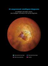
intelligent diagnosis AI intelligent annotation injects new intelligent momentum into fundus diagnosis.
Open the catalog to page 8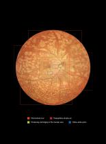
Chorioretinal scar Peripapillary atrophy arc Yellow-white spots Thickening and bulging of the macular area
Open the catalog to page 9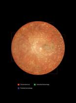
Chorioretinal scar Intraretinal hemorrhage Preretinal hemorrhage
Open the catalog to page 10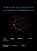
Optic disc analysis ♦ Rim width ♦ Optic disc area ♦ Optic disc tilt rate ♦ Optic disc diameter ♦ Width of the atrophy arc ♦ Height of the atrophy arc ♦ Area of the atrophy arc ♦ Ratio of the area of the atrophy arc to the area of the optic disc ♦ Ratio of the width of the atrophy arc to the width of the optic disc ♦ Arteriovenous diameter ratio ♦ Mean vascular density
Open the catalog to page 11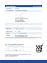
Product model 2/3/4 lasers Imaging technology Confocal laser scanning technology Imaging mode Fundus color photography MC Infrared imaging IR *Red-free imaging RF *Fundus autofluorescence FAF *Fluorescein angiography FFA *Indocyanine green angiography ICGA *Simultaneous angiography FFA + ICGA *Blood flow measurement *Photoreceptor measurement Laser light source Field of View Imaging range Acquisition speed Operation AI analysis Four wavelength light sources: blue, green , red, and infrared >115°, with 7 angles available for selection Single fundus imaging: 170° One-touch fully automatic operation...
Open the catalog to page 12All Big Vision Medical catalogs and technical brochures
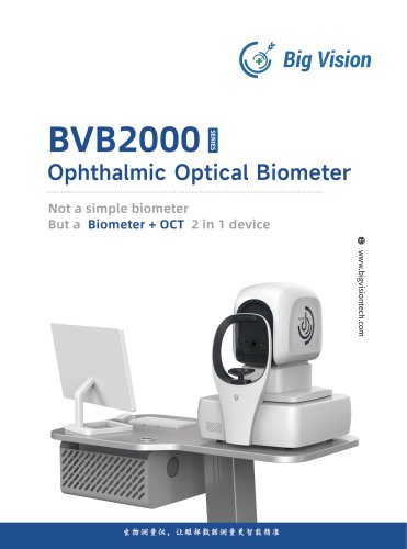 Biometer BVB2000
5 Pages
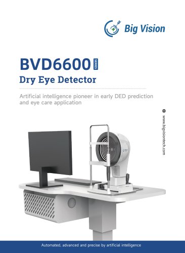 Dry eye BVD6600
8 Pages
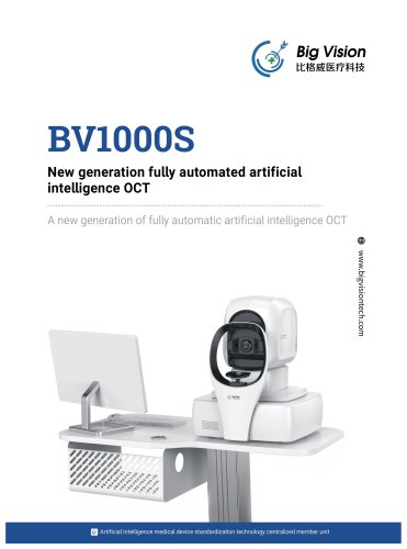 OCT Opthalmoscope BV1000
8 Pages
- Analysis software
- Tablet computer software
- Tablet PC software
- Control software
- Monitoring software
- Diagnostic software
- Fixed ophthalmic examination
- Treatment software
- Data analysis software
- Real-time software
- Ophthalmoscope
- Interpretation software
- Smartphone software
- Screening software
- Retinal camera
- Ophthalmic software
- Ophthalmic biometer
- Eye software
- OCT ophthalmoscope
- Aid software