- Catalogs
- BioActs Co., Ltd
- MitoFlamma® Deep Red (live) technical information
MitoFlamma® Deep Red (live) technical information
1 /1Page
MitoFlamma® Deep Red (live) technical information
1 /1Page
Catalog excerpts
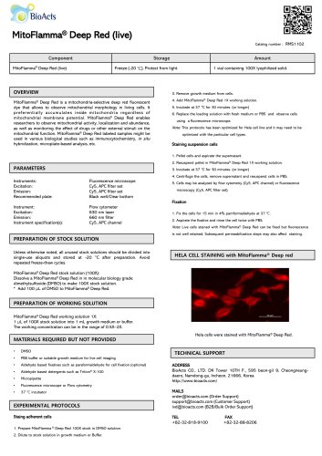
Fluorescence microscope Cy5, APC filter set Cy5, APC filter set Black well/Clear bottom Flow cytometer 630 nm laser 660 nm filter Cy5, APC channel PREPARATION OF STOCK SOLUTION MitoFlamma® Deep Red is a mitochondria-selective deep red fluorescent dye that allows to observe mitochondrial morphology in living cells. It preferentially accumulates inside mitochondria regardless of mitochondrial membrane potential. MitoFlamma® Deep Red enables researchers to observe mitochondrial activity, localization and abundance, as well as monitoring the effect of drugs or other external stimuli on the mitochondrial function. MitoFlamma® Deep Red labeled samples might be used in various biological studies such as immunocytochemistry, in situ hybridization, microplate-based analysis, etc. Recommended plate: Instrument specification(s): 3. Remove growth medium from cells. 4. Add MitoFlamma® Deep Red 1X working solution. 5. Incubate at 37 °C for 30 minutes. (or longer) 6. Replace the loading solution with fresh medium or PBS and observe cells using a fluorescence microscope. Note: This protocols has been optimized for Hela cell line and it may need to be optimized with the particular cell types. Staining suspension cells 1. Pellet cells and aspirate the supernatant. 2. Resuspend pellet in MitoFlamma® Deep Red 1X working solution. 3. Incubate at 37 °C for 30 minutes. (or longer) 4. Centrifuge the cells, remove supernatant and resuspend cells in PBS. 5. Cells may be analyzed by flow cytometry (Cy5, APC channel) or fluorescence microscopy (Cy5, APC filter set). 2. Aspirate the fixative and rinse the cell twice with PBS. Note. Live cells stained with MitoFlamma® Deep Red can be fixed but fluorescence is not well retained. Subsequent permeabilization steps may also affect staining. PREPARATION OF WORKING SOLUTION Unless otherwise noted, all unused stock solutions should be divided into single-use aliquots and stored at -20 °C after preparation. Avoid repeated freeze-thaw cycles. MitoFlamma® Deep Red stock solution (100X): Dissolve a MitoFlamma® Deep Red in in molecular biology grade dimethylsulfoxide (DMSO) to make 100X stock solution. * Add 100 pL of DMSO to MitoFlamma® Deep Red. MitoFlamma® Deep Red working solution 1X: 1 pL of 100X stock solution into 1 mL growth medium or buffer. The working concentration can be in the range of 0.5X-2X. HELA CELL STAINING with MitoFlamma® Deep red MATERIALS REQUIRED BUT NOT PROVIDED Hela cells were stained with MitoFlamma® Deep Red. EXPERIMENTAL PROTOCOLS TECHNICAL SUPPORT • PBS buffer or suitable growth medium for live cell imaging • Aldehyde based fixatives such as paraformaldehyde for cell fixation (optional) • Aldehyde based detergents such as Trition® X-100 • Fluorescence microscope or Flow cytometry Staing adherent cells BioActs CO., LTD. DK Tower 10TH F., 595 beon-gil 9, Cheongneung- daero, Namdong-gu, Incheon, 21666, Korea [email protected] (Order Support) [email protected] (Customer Support) [email protected] (B2B/Bulk Order Support) 1. Prepare MitoFlamma® Deep Red 100X stock in DMSO solution. 2. Dilute to stock solution in growth medium or
Open the catalog to page 1