
- Catalogs
- BioHorizons
- laser-lok

laser-lok

laser-lok
Laser-Lok microchannels are a proprietary dental implant surface treatment developed by BioHorizons. This technology uses laser ablation to create precise microchannels that enhance the attachment and organization of osteoblasts and fibroblasts, promoting tissue growth and a biologic seal around the implant.
The Laser-Lok surface features a consistent microstructure and nanostructure that maximizes surface area, allowing cell pseudopodia and collagen microfibrils to interdigitate with the implant surface. This design inhibits epithelial downgrowth and supports connective tissue attachment, maintaining crestal bone health.
Studies have shown that Laser-Lok implants reduce bone loss significantly compared to traditional implant surfaces. For instance, a multi-center study demonstrated a 70% reduction in bone loss at 37 months post-operation. The technology is effective in various site conditions and has been shown to outperform other designs like NobelReplace™ Select in maintaining bone levels.
Unlike grit-blasted or acid-etched surfaces, which are random and vary in cell reaction, the Laser-Lok surface is consistent and effective for both osseointegration and soft tissue attachment. This has been confirmed through various microscopy techniques.
Recent studies focus on applying Laser-Lok technology to abutments, creating a biologic seal and enhancing peri-implant health. This approach has shown potential in regenerating crestal bone and achieving zero bone loss, which is a future goal for BioHorizons.
Numerous studies have been conducted to evaluate the effectiveness of Laser-Lok technology. These include assessments of immediate or delayed placement and loading, single-tooth replacements, and the impact of laser microtexturing on crestal bone levels. Results consistently show improved bone maintenance and soft tissue integration.
Laser-Lok technology represents a significant advancement in dental implant surfaces, offering superior tissue attachment and bone preservation. Ongoing research continues to explore its full potential in clinical applications.
The document discusses various studies on the impact of Laser-Lok® microtexturing on dental implants, focusing on clinical attachment levels and crestal bone remodeling. The primary aim is to evaluate the effectiveness of Laser-Lok® surfaces in improving implant outcomes.
Materials and Methods: A prospective, randomized study with 77 patients divided into two groups: control (non-Laser-Lok®) and test (Laser-Lok®). Clinical parameters such as crestal bone loss (CBL), clinical attachment level (CAL), plaque index (PI), and bleeding on probing (BOP) were measured at baseline and at 6, 12, and 24 months.
Results: Both groups had a survival rate of 96.1% after 2 years. The Laser-Lok® group showed significantly less CAL loss (0.56mm) and CBL (0.49mm) compared to the control group.
Conclusions: Laser-Lok® implants resulted in better CAL and reduced CBL, although survival rates were similar.
Objectives: To assess clinical, radiographic, and esthetic outcomes of Laser-Lok® implants in the anterior maxilla.
Methods: 46 implants were placed and restored immediately in patients with specific gingival conditions. Evaluations were conducted at 6, 12, and 24 months.
Results: A survival rate of 95.6% was observed. Marginal bone loss was minimal, and soft tissue levels showed slight recession.
Methods: 13 implants were placed with bone regenerative procedures. Evaluations focused on tissue attachment and bone loss.
Results: Laser-Lok® collars provided favorable conditions for tissue attachment and reduced bone loss.
Methods: Examined the effects of laser microgrooves on connective tissue attachment in dogs.
Results: Laser microgrooves facilitated connective tissue attachment, preventing crestal bone resorption.
Introduction: Presented human histology of connective tissue integration with laser microgrooved abutments.
Conclusion: Demonstrated successful connective tissue integration with functionally oriented fibers.
Overall, the studies indicate that Laser-Lok® microtexturing enhances connective tissue attachment and reduces bone loss, offering improved outcomes for dental implants.
This document discusses the impact of laser microtexturing on dental implants, focusing on the Laser-Lok (LL) surface treatment. The LL treatment aims to enhance bone and connective tissue attachment while inhibiting epithelial downgrowth. Various studies and case reports are presented to evaluate the efficacy of LL implants compared to traditional machined implants.
The LL implants feature a 2 mm wide collar with laser micromachining in the lower 1.5 mm. This design is intended to promote osseointegration and connective tissue attachment. The document includes data from a multicenter clinical trial involving 20 pairs of implants in 15 patients, with measurements taken over 37 months.
1. Clinical Trials: LL implants showed significantly less crestal bone loss and shallower probing depths compared to control implants. At 37 months, LL implants had a mean probing depth of 2.30 mm and crestal bone loss of 0.59 mm, compared to 3.60 mm and 1.94 mm, respectively, for control implants.
2. Comparative Studies: LL implants demonstrated superior performance in maintaining crestal bone levels and soft tissue attachment compared to Nobel Replace Select implants. LL implants showed less crestal bone loss and shallower probing depths at both 6 and 12 months.
3. Long-term Case Studies: Case reports highlighted the ability of LL implants to maintain crestal bone levels and soft tissue esthetics over extended periods, with some cases showing stability up to 13 years post-restoration.
The consistent performance of LL implants in reducing crestal bone loss and maintaining soft tissue attachment suggests a stable soft-tissue seal above the crestal bone. The LL design may redefine the "normal" implant biologic width by providing a supracrestal connective tissue attachment. The studies support the clinical relevance of establishing a biologic seal of connective tissue fibers around dental implants.
Figures in the document illustrate the dimensions of healing abutments, implant placement procedures, and comparative data on crestal bone loss and probing depths. Key data points include:
- LL implants showed a mean crestal bone loss of 0.44 mm at 2 years and 0.46 mm at 3 years post-restoration.
- LL implants maintained crestal bone levels within 0.5 mm of the implant/abutment interface in long-term studies.
The document recommends considering LL implants for their ability to preserve crestal bone levels and enhance soft tissue attachment. The "one abutment, one time" approach is suggested to minimize tissue changes during the healing phase.
This document reviews various studies on the effects of laser-ablated microgrooves on dental implants, focusing on their impact on bone and soft tissue attachment, as well as stress distribution in the surrounding bone.
The studies involved different implant designs, including microgrooved and turned collar implants, and examined their effects on bone-to-implant contact (BIC) and marginal bone loss. The research utilized finite element analysis (FEA) to assess stress distribution and histomorphometric analysis to evaluate tissue response.
- Microgrooved implants showed higher BIC and reduced marginal bone resorption compared to turned collar implants.
- Laser microgrooves inhibited apical migration of crevicular epithelium and promoted true gingival attachment.
- FEA indicated that sub-crestal placement of implants reduced stress and strain in surrounding bone.
- Laser-Lok implants demonstrated decreased crestal bone loss and advantageous tissue response compared to other designs.
- Figures illustrated histological sections and stress distribution models, highlighting differences in tissue attachment and stress levels between implant types.
- Data showed that laser microgrooved implants had significantly lower stress levels and better bone integration compared to control implants.
Laser-ablated microgrooves on implant collars provide favorable conditions for hard and soft tissue attachment, reducing marginal bone resorption and enhancing implant stability. The studies suggest that these implants may improve long-term performance by mimicking natural tooth attachment mechanisms.
The studies were limited by their pre-clinical nature and the specific animal models used, which may not fully replicate human clinical conditions.
Tissue response to implantable devices is influenced by material interface parameters such as composition, surface chemistry, and microgeometry. Surface microgeometry plays a significant role in tissue-implant interactions, with rough surfaces promoting bone integration and smooth surfaces leading to fibrous tissue encapsulation. Textured surfaces enhance mechanical stability and influence cell adhesion and growth.
Studies have shown that controlled surface microgeometry can enhance bone integration and influence fibroblast colonization. Titanium implants with laser-ablated microstructures were tested, showing that specific microgeometry sizes can improve bone integration.
Research on fibroblast variants cultured on micropatterned surfaces revealed that surface geometry affects cell shape, orientation, and cytoskeletal organization. Cells on grooved surfaces aligned with the grooves, while those on posted surfaces showed stress fibers terminating on posts. These findings suggest that microgeometry can direct tissue response and improve implant integration.
Surface microgeometry influences fibroblast shape and cytoskeletal organization, affecting cell growth and behavior. Grooved surfaces require specific depth and width to influence cell orientation and growth direction. Rough surfaces promote thinner fibrous capsules, enhancing implant integration.
Surface microgeometry plays a crucial role in tissue-implant interactions, affecting cell behavior and implant integration. Further research into phenotypic differences and extracellular matrix roles could lead to improved implant designs and longer functional lifespans.
This document provides an overview of various studies and references related to dental implants, specifically focusing on the effects of laser microtexturing on implant collars, crestal bone levels, and peri-implant health. It also includes warranty and product information for BioHorizons dental products.
- Radiographic Analysis of Crestal Bone Levels: This study examines the impact of Laser-Lok® collar dental implants on crestal bone levels, highlighting the benefits of laser microtexturing in maintaining bone health.
- Effects of Laser Microtexturing: Research by Botos et al. explores how laser microtexturing affects crestal bone levels and peri-implant health, suggesting improved outcomes with this technology.
- Marginal Tissue Response: Bae et al. investigate different implant neck designs and their influence on tissue response, emphasizing the importance of design in implant success.
- Lifetime Warranty: BioHorizons offers a lifetime warranty on implants and prosthetic components, covering failures excluding normal wear.
- Additional Warranties: Surgical drills, taps, and instruments have specific warranty periods, with recommendations for replacement based on usage and wear.
- Return Policy: Products can be returned with a Return Authorization Form, and the use of non-BioHorizons components voids warranties.
BioHorizons emphasizes the responsibility of clinicians in treatment planning and product use, recommending postgraduate education and adherence to instructions for use. The company limits its liability to product replacement or repair under warranty conditions.
BioHorizons products are available in various countries, with compliance to international standards such as ISO 13485:2003. The document notes that not all products are available in all regions, and specifications may change.
Contact details for BioHorizons offices in multiple countries are provided for customer support and inquiries.
Catalog excerpts
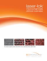
The dental implant surface that elicits a true connective tissue attachment SCIENCE • INNOVATION • SERVICE
Open the catalog to page 1
Laser-Lok Technology SEM image at 39X showing the Laser-Lok zone on a BioHorizons Tapered Plus implant. Laser-Lok at 800X exhibits consistently formed microchannels to organizeand promote tissue growth.2,3,4,5,6,7,8,9,10,16,17 The uniformity of the Laser-Lok microstructure and nanostructure is evident using extreme magnification. Laser-Lok overview Laser-Lok microchannels is a proprietary dental implant surface treatment developed from over 25 years of research initiated to create the optimal implant surface. Through this research, the unique Laser-Lok surface has been shown to elicit a biologic...
Open the catalog to page 2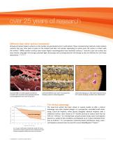
over 25 years of research Different than other surface treatments Virtually all dental implant surfaces on the market are grit-blasted and/or acid-etched. These manufacturing methods create random surfaces that vary from point to point on the implant and alter cell reaction depending on where each cell comes in contact with the surface.10 While random surfaces have shown higher osseointegration than machined surfaces,11 only the Laser-Lok surface has been shown using light microscopy, polarized light microscopy and scanning electron microscopy to also be effective for soft tissue attachment.2,3,4,5,6,7,8,9,10...
Open the catalog to page 3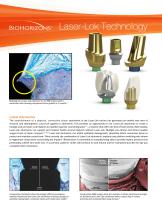
Laser-Lok Technology Histology of a Laser-Lok abutment on an RBT implant with a machined collar showing exceptional bone growth at 3 months.6 Latest discoveries The establishment of a physical, connective tissue attachment to the Laser-Lok surface has generated an entirely new area of research and development: Laser-Lok applied to abutments. This provides an opportunity to use Laser-Lok abutments to create a biologic seal and Laser-Lok implants to establish superior osseointegration15 – a solution that offers the best of both worlds. Alternatively, Laser-Lok abutments can support peri-implant...
Open the catalog to page 4
Latest Implant Research Challenging the Tarnow rule with Tapered Plus Study Review The following review summarizes many of the studies and presentations related to Laser-Lok microchannels. Latest Abutment Research Laser-Lok abutment proof of principle (canine) 10 Laser-Lok ball abutment case report (human) 11 Laser-Lok abutment case series (human) 12 Laser-Lok abutment tissue reattachment (human) 13 Reusing Laser-Lok abutments (canine) 14 Additional Human Studies Additional Pre-clinical Research Three year prospective, controlled
Open the catalog to page 5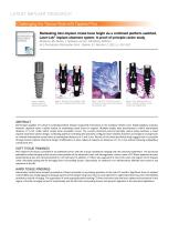
LATEST IMPLANT RESEARCH Challenging the Tarnow Rule with Tapered Plus Maintaining inter-implant crestal bone height via a combined platform-switched, Laser-Lok® implant/abutment system: A proof-of-principle canine study. M Nevins, ML Nevins, L Gobbato, HJ Lee, CW Wang, DM Kim. Int J Periodontics Restorative Dent , Volume 33, Number 3, 2013. p. 261-267. Figure 1. Platformswitched Laser-Lok abutment and implant Figure 2. No signs of inter-implant crestal bone loss on implants placed 2mm apart Figure 3. Direct CT connection to LaserLok surfaces; osseous crest extends onto laser-ablated collar Figure...
Open the catalog to page 6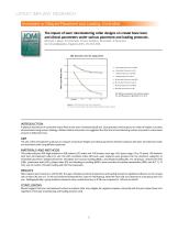
LATEST IMPLANT RESEARCH Immediate or Delayed Placement and Loading, Controlled The impact of laser microtexturing collar designs on crestal bone level, and clinical parameters under various placement and loading protocols. M Serra, L Bava, D Farronato, V Iorio Siciliano, M Grande, R Guarnieri. Int J Oral Maxillofac Implants 2014; 29:354-363. CBL Outcomes over the study period 0 Bone Loss Mean (mm) -0.2 L Immediate Placement and INOL L Immediate Placement and DL L Delayed Placement and INOL L Delayed Placement and DL M Immediate Placement and INOL M Immediate Placement and DL M Delayed Placement...
Open the catalog to page 7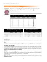
Immediate occlusal loading of tapered internal Laser-Lok implants in partial arch application: A 24-months clinical and radiographic study. M Grande, A Ceccherini, M Serra, L Bava, D Farronato, V lorio Siciliano, R Guarnieri. J Osseointegr 2013;5(2):53-60. BACKGROUND Recently, new implant surfaces have been proposed in an effort to improve hard and soft tissue integration, which may be beneficial in immediate loading situations. AIM The purpose of the present prospective clinical study was to, during 2 years, clinically and radiographically evaluate an implant with laser microtextured collar...
Open the catalog to page 8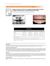
Influence of Laser-Lok® surface on immediate functional loading of implants in single tooth replacement: a 2-years prospective clinical study. D Farronato, F Mangano, F Briguglio, V lorio Siciliano, R Guarnieri. Int J Periodontics Restorative Dent, Volume 34, Number 1. p. 79-89. Figure 1. Orthopantomography of the patient who received Figure 2. Before treatment (A), after treatment (B) two implants (1 LL in site 14 and 1N LL in site 24) Mean values and SD t=2.7338, p=0.0340, the difference between the observed means is significant for p <0.05 Figure 3. CBL outcomes over the study period ABSTRACT...
Open the catalog to page 9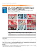
Clinical, Radiographic, and Esthetic Evaluation of Immediately Loaded Laser Microtextured Implants Placed into Fresh Extraction Sockets in the Anterior Maxilla: A 2-Year Retrospective Multicentric Study Guarnieri, Renzo MD, DDS; Placella, Roberto DDS; Testarelli, Luca DDS, PhD; iorio-Siciliano, Vincenzo DDS, PhD; Grande, Maurizio DDS implant Dentistry, April 2014, Volume 23, issue 2, p. 144-154. Table 1. Marginal Bone loss in Relation to BSL at T1 (6 months), T2 (1 year), and T3 (2 years) OBJECTIVES To assess the clinical, radiographic, and esthetic outcomes of implants with a laser microtextured...
Open the catalog to page 10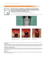
LATEST IMPLANT RESEARCH Single-stage Implants with LADDEC® and Mem-Lok® Soft and hard tissue modifications at immediate transmucosal implants (Laser-Lok® microtextured collar) placed into fresh extraction sites. A six month prospective study with surgical re-entry. V Iorio Siciliano, G Marzo, A Blasi, C Cafiero, M Mignogna, M Nicolò Int J Periodontics Restorative Dent, Volume 34, Number 4, 2014. p. 541–549. Figure 1. Schematic of the Single-stage Implant System. Figure 2. Clinical view after filling of the peri-implant defects with bone-derived xenograft particles (Laddec® BioHorizons, IPH. Inc)....
Open the catalog to page 11All BioHorizons catalogs and technical brochures
 International Product Catalog
12 Pages
 Biologics Catalog
28 Pages
 External Dental Implant Catalog
44 Pages
 patientconnect
24 Pages
 prosthetics catalog 2017
36 Pages
 External Dental Implant
44 Pages
 Surgical Motors
2 Pages
 AlloDerm ®
2 Pages
 biologics product catalog
12 Pages
 one-piece 3.0 & overdenture
28 Pages
 laser-lok 3.0
20 Pages
 Hu-Friedy® dental instruments
24 Pages
 guided surgery kit
24 Pages
 tapered internal implant system
28 Pages


Archived catalogs
 prosthetics catalog
40 Pages
- Implant abutment
- Titanium implant abutment
- Straight implant abutment
- Dental surgery instrument kit
- Dental burr
- Titanium dental implant
- Conical dental implant
- Dental implant surgery instrument kit
- Bone substitute
- Cylindrical dental implant
- Dental implant analog
- Straight dental implant analog
- Hexagonal dental implant
- Narrow dental implant
- Internal hexagon dental implant
- Titanium healing abutment
- Impression coping
- Dental sugery bone substitute