- Catalogs
- Biolase Tech.
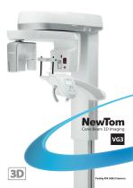
- NewTom VG3
NewTom VG3

NewTom VG3
NewTom Cone Beam 3D imaging units, developed by QR s.r.l. in Italy, are leaders in dental imaging technology. The NewTom 9000, introduced in 1996, was the first Cone Beam system and set the standard for future models. These units are known for their reliability and advanced technology.
Specifications and Features
The Cone Beam 3D imaging technology provides a large field of view (FOV) to capture the entire mandible and maxilla, crucial for comprehensive diagnosis. It uses a cone-shaped beam for a single rotation scan, reducing radiation exposure compared to traditional CT scans. SafeBeam™ technology further minimizes radiation by adjusting dosage based on the density of the scanned volume.
Software and Compatibility
NewTom's NNT software supports 2D and 3D image reconstruction and analysis, offering high-quality imaging for various dental applications. It is compatible with major third-party programs, enhancing its utility in diagnosis, treatment planning, and patient education. The NewTom Implant Planning software allows for detailed 3D implant simulations and surgical planning.
Applications and Benefits
Cone Beam 3D imaging is essential for implantology, orthodontics, periodontics, and oral/maxillofacial surgery. It provides precise 1:1 scale imaging, crucial for accurate diagnosis and treatment planning. The technology supports multiple FOVs, allowing tailored scans for specific diagnostic needs while adhering to ALARA principles to minimize radiation exposure.
Patient Comfort and Safety
NewTom systems are designed to enhance patient comfort and reduce movement during scans, improving image quality. The systems produce high-resolution images with minimal radiation exposure, contributing to better patient acceptance and understanding of treatment plans.
Conclusion
NewTom Cone Beam 3D imaging systems represent a significant advancement in dental imaging technology, offering high-quality, low-radiation imaging solutions compatible with a wide range of diagnostic and treatment planning software.
Overview of NewTom VG3
The NewTom VG3 is an advanced imaging device for dental specialists, offering a range of 2D and 3D examinations. It is particularly useful for evaluating impacted teeth, fractures, and bone irregularities, and is adaptable for various dental applications including dentures, braces, and implants.
2D Examinations
The device supports multiple 2D imaging options such as bite wings, panoramic, sinuses, TMJ, cephalometric radiographs, and carpal radiography. Cephalometric radiographs are crucial for diagnosing facial growth abnormalities and planning orthodontic interventions. Carpal radiography helps assess bone growth in children.
Benefits
The NewTom VG3 features the largest Field of View (FOV) available, combined with high CBCT technology for clear images. It includes SafeBeam™ Technology to adjust radiation dosage for patient safety and offers multiple FOV and scan modes. The device is user-friendly with NNT software for easy image sharing and can be upgraded to 3D and Ceph configurations.
Technical Specifications
The NewTom VG3 uses a high-frequency, stationary anode X-ray source with a 0.5 mm focal spot. It features a flat panel amorphous silicon detector with a resolution of 10.4 lp/mm. The device supports single scan and Cone Beam acquisition, with a dynamic range of 16-bit. It offers various FOV sizes and voxel size options, with patient positioning options for standing or seated, including wheelchair accessibility.
Design and Usability
The NewTom VG3 is compact, does not require an air-conditioned room, and is easy to install without complex radiation protection structures. It includes a touch screen panel, USB port, and a user-friendly menu, making it a practical choice for dental practices.
Power and Dimensions
The device requires 15A @ 115V or 10A @ 240V power supply. It has adjustable height and width dimensions, with a total weight of 170 Kg, making it suitable for various clinical settings.
Catalog excerpts
Cone Beam 3D Imaging
Open the catalog to page 1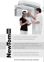
Large FOV to include the entire mandible and maxilla for a complete diagnosis. Latest technology utilized to create perfect panoramic images with an accurate focal trough over the whole arch. New generation of the NNT Software for all types of image reconstructions and analyses. First in Cone Beam, Accurate in Results
Open the catalog to page 2
FIRST USER OF CONE BEAM IN DENTAL FIELD QR s.r.l. is the name that stands behind NewTom Cone Beam 3D imaging units and the creator of Cone Beam technology for the dental field. The NewTom 9000 (also known as Maxiscan) was the first Cone Beam system in the world and was originally installed in 1996. This unit was the forefather of the NewTom product line and of all X-Ray units based on Cone Beam technology. QR’s 20 plus years of experience and success in research, development, manufacturing and distribution of NewTom products affirm our commitment to excellence and quality. QR s.r.l. is based...
Open the catalog to page 3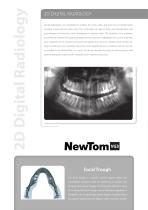
2D DIGITAL RADIOLOGY Dental radiography was developed to visualize the entire upper and lower jaws including teeth, maxillary sinuses and the nasal cavity. This technology can help to study and evaluate bone and gum diseases, jaw fractures, tooth development, impacted teeth, TMJ disorders, sinus problems and other oral diseases. Principal advantages are that panoramic radiographs can cover a large area, give a bilateral view of anatomy and expose the patient to a minimum radiation dose thereby making the treatment more tolerable. One of the most valuable features is however that the unit can...
Open the catalog to page 4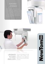
REMOVABLE 2D SENSOR NewTom increase the value of this system by adding removable sensor technology. This allows the operator to safely switch the 2D sensor from the main structure and use it on the Ceph arm. A removable 2D sensor is the perfect solution for those practices which require a high quality device at a competitive price. Through the development of a truly modular system, NewTom VG3’s performances can be upgraded to CB3D and Ceph with minimal effort, at any time.
Open the catalog to page 5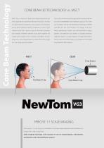
MSCT uses a narrow fan beam that rotates around the pa- The result is a more accurate image without missing informa- tient acquiring thin axial slices with each revolution. In order tion and a considerably lower radiation exposure. The Ame- to create a section of anatomy, many rotations must be done. rican Academy of Oral and Maxillofacial Radiology (AAOMR) During these repeated rotations, traditional CT emits a high prescribes the use of Cone Beam 3D imaging when eva- radiation dose, but it leaves a gap of information between luating periodontal, implant, and oral/maxillofacial surgery each...
Open the catalog to page 6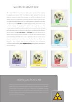
MULTIPLE FIELDS OF VIEW The scanner’s FOV determines how much of the patient’s anatomy will be visualized. If using a flat panel detectors (FPD), the dimensions of their cylindrical FOV can be described as diameter by height (DxH). Nowadays, the need to scan different R.O.I. with different dimensions is regulated by international standards in order to reduce the effective dose to the patient following the “As Low As Reasonably Achievable” (ALARA) dose principles. The use of a small FOV (on user-defined region in endo, perio, implant surveys and for the localization of impacted teeth) in addition...
Open the catalog to page 7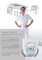
SAFEBEAM™ TECHNOLOGY FOR AUTOMATIC DOSE EXPOSURE Only NewTom Cone Beam systems employ SafeBeam™ technology, the safest technology available for patient and staff. Featured in all NewTom units, SafeBeam™ automatically adjusts the radiation dosage according to the density of the volume in the gantry. This technology uses intermittent bursts of radiation, which last only milliseconds, during image acquisition. Other systems deliver a constant stream of radiation and the same amount of radiation, whether scanning a 100 Kg adult or a small child. SafeBeam™ technology automatically and continuously...
Open the catalog to page 8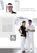
GREATER PATIENT COMFORT AND TREATMENT ACCEPTANCE All NewTom units add a sense of comfort for the patients, allowing them to relax during the scan and limiting the patient movements, in order to improve the image quality. NewTom scans provide the practitioner and the patient with unprecedented visualization of cranial anatomic information. This leads to a better diagnosis and better treatment planning, increasing the patient treatment knowledge. The result is a more cooperative and informed consent process along with understanding the need for treatment and improving the doctor-patient relationship....
Open the catalog to page 9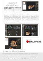
NEWTOM NNT ANALYSIS SOFTWARE NewTom NNT analysis software is the perfect solution for 2D and 3D imaging. NNT allows the creation of different kinds of 2D and 3D images in a 16 bit grey-scale and it takes only few seconds to evaluate the data taken during the scan. The software is totally designed by NewTom engineers and, thanks to the various application modes specifically design for can be delivered digitally (burnt to a CD or DVD), on paper, different fields of use, it fulfills all the requirements and needs film or pdf. The software is available in different versions: the of our clients. NNT,...
Open the catalog to page 10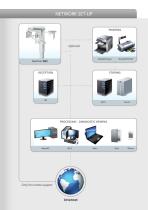
NETWORK SET-UP PRINTING DICOM Printer Standard Printer PROCESSING - DIAGNOSTIC VIEWING Only for remote support
Open the catalog to page 11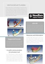
NEWTOM IMPLANT PLANNING NewTom Implant Planning is a software package which allows the creation of 3D implant simulation. The software can simulate the implant placement on 2D and 3D models, identify the mandibular canal along with drawing panoramic and cross sections of the bone model. It also shows the 3D bone model and calculates the bone density. The NewTom Implant Planning software is used to plan prosthesis implant surgery in a faster, safer and more efficient way. It also allows the ability to export in .stl format. 2D & 3D The NIP software generates beautiful panoramic images, cross sections...
Open the catalog to page 12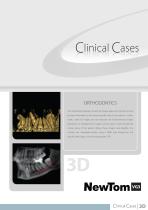
Clinical Cases ORTHODONTICS The combination between 2D and 3D images allows the clinician to have the best information at the lowest possible dose to the patient. In other words, when 3D images are not necessary the bi-dimensional images (panoramic or cephalometric images) can be used to show the general clinical status of the patient. Where those images look doubtful, the clinician can investigate further using a CB3D scan focused on the specific pathology using the appropriate FOV.
Open the catalog to page 13All Biolase Tech. catalogs and technical brochures
 Epic
Epic1 Page
 EPIC-US Brochure
2 Pages
 NewTom VGI
13 Pages
 GALAXY BioMill CAD/CAM
2 Pages
 iLase 2012 brochure
2 Pages
 iPlus
iPlus6 Pages

