
- Catalogs
- Biolase Tech.
- NewTom VGI

NewTom VGI

NewTom VGI
NewTom is a leader in Cone Beam 3D imaging technology, offering dental imaging solutions that prioritize reduced radiation exposure and advanced technology. Their products are manufactured in Italy, focusing on reliability and innovation.
Specifications and Features
NewTom's Cone Beam 3D imaging systems provide medical-grade performance with features like a rotating anode, adjustable field of view (FOV), and rapid image reconstruction. The SafeBeam™ technology minimizes radiation doses, with exposure times under 4 seconds. The system is designed for patient comfort, including wheelchair accessibility and a compact design.
Comparison with Traditional Imaging
Cone Beam 3D imaging captures complete images in a single rotation, unlike traditional CT scans that require multiple rotations and emit higher radiation. This technology is recommended for dental evaluations, including periodontal and maxillofacial surgeries, due to its accuracy and lower radiation.
Field of View (FOV) and Radiation Dose
The FOV determines the anatomical area visualized, with options for different sizes to minimize radiation exposure according to ALARA principles. Smaller FOVs enhance image resolution, while larger FOVs cover extensive anatomical regions.
Software and Compatibility
NewTom's NNT software supports the creation of 2D and 3D images, aiding detailed analysis and surgical planning. It is compatible with major third-party applications, enhancing its utility in diagnostics and education.
Clinical Applications
Cone Beam 3D imaging is essential for implantology, orthodontics, and oral/maxillofacial surgery, providing detailed anatomical insights and aiding in precise treatment planning. It supports accurate implant assessments and TMJ evaluations.
Conclusion
NewTom's Cone Beam 3D imaging systems offer a comprehensive solution for dental professionals, combining high-resolution imaging with patient safety and comfort. Their advanced software and compatibility with third-party applications make them a versatile tool in modern dental practices.
NewTom VGi Overview
The NewTom VGi is a Cone Beam Computed Tomography (CBCT) scanner used in dental and ENT fields, known for its technological advantages.
Specifications
The NewTom VGi features a high-frequency X-ray source with a rotating anode, operating at 110 kV and 1-20 mA in pulsed mode. It has a focal spot of 0.3 mm and uses SafeBeam™ control to reduce radiation based on patient size. The effective dose is 99 μSv for a full FOV, with scan times of 18s/26s and X-ray emission time of 3.6s in standard mode. It uses an amorphous silicon flat panel detector with a 20 cm x 25 cm field of view.
Applications
Endo-Perio: Provides high-quality images for diagnosing tooth structure and pathology.
ENT Protocols: Considered the gold standard for ENT examinations, offering detailed imaging with minimal radiation.
Orthodontics: Produces accurate 3D images for cephalometric tracings and assessing impacted teeth.
Benefits
The NewTom VGi offers clear images with a small focal spot, enhanced patient comfort, and reduced radiation exposure. It features multiple FOV options and scan modes, and is equipped with NNT software for easy image sharing. The scanner is wheelchair accessible and does not require a specialized room setup.
Functioning and Training
The scanner operations are computer-guided with user-friendly menus, supporting the user through each step of the scanning process. It includes a viewer and sharing application, is DICOM conformant, and offers improved software integration.
Conclusion
The NewTom VGi represents advanced CBCT technology, providing high-quality imaging with reduced radiation exposure, suitable for various medical and dental applications.
Catalog excerpts
Cone Beam 3D Imaging Exclusive Dental Distributor for North America
Open the catalog to page 1
Cone Beam 3D Imaging True medical grade imaging technology at a fraction of the cost and radiation exposure
Open the catalog to page 2
Pioneers of Cone Beam in the Dental Field QR s.r.l. is the name that stands behind NewTom Cone Beam 3D imaging units and we were the creators of Cone Beam technology for the dental field. NewTom 9000 (also known as Maxiscan) was the very first Cone Beam in the world, which was installed in 1996. It pioneered the NewTom product line and, in general, the entire X-Ray units based on Cone Beam technology. QR’s 20 plus years of experience and success distribution of NewTom products afirms our commitment to excellence and quality. QR s.r.l. is based in Italy and all NewTom products are designed and...
Open the catalog to page 3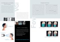
Cone Beam 3D vs. CT Imaging and 2D Imaging The scanner’s FOV determines how much of the patient’s anatomy biggest FOV (which include the roof of the orbits and the Nasion Traditional CT (CAT scan) uses a narrow fan beam that rotates will be visualized. If using a flat panel detector (FPD), the dimensions down to the hyoid bone) permits with one single rotation to around the patient acquiring thin axial slices with each revolution. In of their cylindrical FOV can be described as Diameter by Height scan patients where the referring doctors need to see the major order to create a section of anatomy,...
Open the catalog to page 4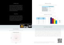
Effective Dose Table: effective dose from conventional dental imaging techniques in Sv. MSCT = multislice CT* Proper assessment for implants requires the visualization of all aspects of the mandibular canal. The ability to see small anato- mical parts such as tooth roots and periodontal ligaments, as Intraoral radiograph Panoramic radiograph quality and the quantity of details necessary to accurately view Cephalometric radiograph the canal for secure implant assessment. MSCT maxillo-mandibular well as any present lesions, is critical in determining successful placement. Only 3D High Resolution...
Open the catalog to page 5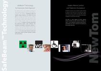
SafeBeam™ Technology for Automatic Dose Exposure Greater Patient Comfort and Treatment Acceptance Only NewTom systems employ SafeBeam™ technology, the All NewTom units add a sense of comfort for patients, allowing safest technology available for patient and staff. Featured in the patient to relax during the scan and limiting the patient all NewTom units, SafeBeam™ automatically adjusts the movements in order to improve the image quality. NewTom radiation dosage according to the patient’s age and scans provide the practitioner and the patient unprecedented size. This technology uses intermittent...
Open the catalog to page 6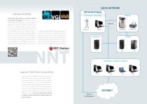
LOCAL NETWORK QR Standard Supply Software Flexibility ACQUISITION - PROCESSING NewTom NNT analysis software is the perfect integration to Cone Beam 3D imaging. NNT allows the creation of different kinds of 2D and 3D images, in a 16 bit grey-scale, and it takes only few seconds to evaluate the data taken during the scan. It is completely designed by NewTom engineers, and it fits all the requirements and needs of our clients. NNT can easily identify and mark root inclination, position of impacted and supernumerary teeth, absorption, hyperplastic permits data processing, an NNT Viewer; that gives...
Open the catalog to page 7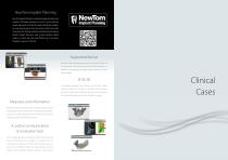
NewTom Implant Planning New Tom Implant Planning is a software package that allows the creation of 3D implant simulation on any PC. It can simulate the implant placement on 2D and 3D models, identify the mandibular canal, draw panoramic and cross sections of the bone model. It also shows the 3D bone model and calculates the bone density. NewTom Implant Planning is used to plan prosthesis implant surgery in a faster, safer and more efficient way. It also allows the ability to export in .stl format. Supported format NewTom Implant Planning reads axials slices saved in DICOM 3.0 or in NNT format,...
Open the catalog to page 8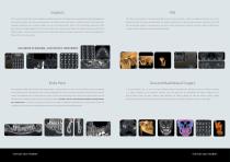
CBCT is one of the most effective tools available for analyzing implant sites. 3D images can accurately identify possible pathologies and CBCT takes the examination of the Temporomandibular Joint to a new level. After a single scan, Sagittal and Coronal views can be structural abnormalities. Cross sectional and panoramic views facilitate various calculations as: height and width of the implant sites, sectioned to show joint space and pathologies. 3D images reconstruction can clearly provide exhaustive information of the TMJ mandibular edentulous site, a potential implant site near the mental...
Open the catalog to page 9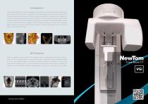
Orthodontics While various pan-cephalometric machines create adequate images, Cone Beam scanners produce many types of images, including panoramic, cephalometric and 3D. Based on the physics of this technology, images are more accurate than 2D dental x-rays and 3D medical scanners. As a result, cephalometric tracings from dental Cone Beam scanners can be generated with confidence. The 3D image, in case of palatal expansion, can clearly show the buccal bone and molar roots in order to avoid unnecessary gingival recession. Impacted teeth may cause dental problems that produce few, if any symptoms....
Open the catalog to page 10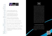
NewTom VGi, from the company that was the first to use the Cone Beam technology in dental field, represents the newest in CBCT technology. NewTom VGi takes an image at every degree of rotation, 360° rotation = 360 images, increasing the range of possibilities for image manipulation. It couples a revolutionary flat panel x-ray detector technology with a very small focal spot (0.3 mm), Clearest images possible with the smallest possible focal spot. to produce the clearest, sharpest images possible. VGi features an adjustable Field Of View, which allows doctors to irradiate just the right volume,...
Open the catalog to page 11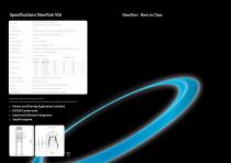
NewTom - Best in Class High frequency, constant potential (DC), X-ray Source rotating anode: 110 kV; 1-20 mA (pulsed mode) Focal Spot X-Ray Cone Beam Proprietary SafeBeam™ control reduces radiation based on patient size. Effective Dose 99 μSv Full FOV (ICRP 2007, estimate for adult) Scan Time X-ray Emission Time Image Acquisition 360 Images - 360 degree rotation Image Detector Amorphous silicon flat panel, 20 cm x 25 cm Field of View (7.87 in x 9.84 in) Signal Grey Scale 14-bit scanning, 16-bit reconstruction FOV sizes D x H Multiples Scan Modes Standard scan Boosted scan HiRes scan Voxel size...
Open the catalog to page 12All Biolase Tech. catalogs and technical brochures
 Epic
Epic1 Page
 EPIC-US Brochure
2 Pages
 NewTom VG3
24 Pages
 GALAXY BioMill CAD/CAM
2 Pages
 iLase 2012 brochure
2 Pages
 iPlus
iPlus6 Pages

