- Catalogs
- Bioquochem
- STAIN LINE CATALOGUE
- Company
- Products
- Catalogs
- News & Trends
- Exhibitions
STAIN LINE CATALOGUE
1 /16Pages

STAIN LINE CATALOGUE
1 /16Pages
Catalog excerpts
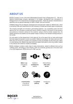
BQCKit Company is one of the three differentiated business lines of Bioquochem S.L. We are a Spanish biotechnology company specialized in the design, development and manufacture of biological kits for the measurement of biological parameters. These kits present characteristics of specificity and are geared especially for fields of health, food and cosmetic. All BQCkit Assay Kits are designed, developed and manufactured in Spain by BQCkit team, which is formed by PhDs and researchers from the University of Oviedo with several years of experience. The company strengths are innovation, personalized...
Open the catalog to page 5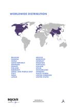
MEXICO MOROCCO NORWAY PAKISTAN PORTUGAL POLAND SPAIN SWITZERLAND SWEEDEN SOUTH KOREA UNITED KINGDOM UNITED STATES BELGIUM CANADA CHINA CZECH REPUBLIC DENMARK FRANCE FINLAND GERMANY ISRAEL AND MIDDLE EAST ITALY INDIA JAPAN www.bqckit.com www.bioquochem.com
Open the catalog to page 6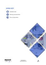
ICON KEY Available sizes Staining characteristics Time of preparation BOOc/t A brand of Qu^fhem
Open the catalog to page 7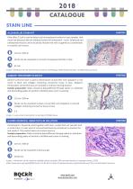
ALCIAN BLUE STAIN KIT Alcian Blue 2.5 pH is used to detect acid mucopolysaccharides in tissue samples, that cannot be detected with the habitual method of hematoxylin – eosin. Sulphured and carboxylated mucins can be visualized. Nuclear fast red is supplied as a counterstain, to visualize cell nucleus. 125 ml / 500 ml Nuclei can be visualized as red and mucopolysaccharides in blue 45 min Image 1 Attribution By The Armed Forces Institute of Pathology (AFIP) [Public domain], via Wikimedia Commons GOMORI TRICHROME STAIN KIT Gomori trichrome stain is used to demonstrate muscle fiber and cytoplasm...
Open the catalog to page 8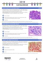
MAYER’S HEMATOXYLIN AND EOSIN STAIN KIT Hematoxylin Eosin is a common staining technique used in a wide range of tissues. Through this method, nuclei can be visualized as blue, muscle in red, connective tissue in pink and nucleic acid associated with proteins in purple. Sample preparation: Slides should be deparaffinized through xylene or substitute and descending grades of alcohol to distilled water prior to staining. 125 ml / 500 ml Nuclei can be visualized as blue, muscle in red, connective tissue in pink and nucleic acid associated with proteins in purple 35-40 min Image 4 Attribution By...
Open the catalog to page 9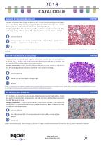
MASSON’S TRICHROME STAIN KIT Masson trichrome stain is used to demonstrate connective tissue elements, collagen and muscle fibers. After staining, collagen and mucin will be coloured in blue, muscle fibers, cytoplasm and keratin in red and the nuclei blue/black. Sample preparation: Formalin fixed, paraffin and frozen samples can be used. After each step, rinsing with tap water and distilled water is required unless specified 125 ml / 500 ml Collagen and mucin can be visualized as blue, muscle fibers, cytoplasm and keratin in red and the nuclei blue/black. 2h Image 7 Attribution: Per Nephron [CC...
Open the catalog to page 10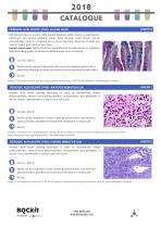
PERIODIC ACID-SCHIFF (PAS)- ALCIAN BLUE PAS-AB technique is used to differentiate between acidic mucins as sialomucins, sulfomucin and neutral epithelial mucin. After staining, acidic mucins will be coloured blue, neutral in magenta and nuclei in deep blue. Mixtures of acidic and neutral mucins give blue/ purple colour. Sample preparation: Slides should be deparaffinized through xylene or substitute and descending grades of alcohol to distilled water prior to staining. 125 ml / 500 ml Acidic mucins can be coloured in blue, neutral in magenta and nuclei in deep blue. Mixtures of acidic and neutral...
Open the catalog to page 11
SCHIFF REAGENT SOLUTION Schiffs reagent is used in the staining technique PAS. Periodic Acid Schiff is used to demonstrate simple polysaccharides, neutral muco-polisaccharides, mucoproteins, and glicolipids. It can be used together with hematoxylin or alcian blue. 125 ml / 500 ml Only used together with the PAS Stain 60 min Image 13 Attribution By The Armed Forces Institute of Pathology (AFIP) [Public domain], via Wikimedia Commons CRESYL VIOLET SOLUTION 0.1 % Cresyl Violet 0,1% is used to demonstrate Nissl substance in the nervous system, X chromatin, Helicobacter, mastocytes and cartilage granules....
Open the catalog to page 12
CRESYL VIOLET SOLUTION 2 %l KH07016 Cresyl Violet 0,1% is used to demonstrate Nissl substance in the nervous system, X chromatin, Helicobacter, mastocytes and cartilage granules. Neurons are visualized in purple and cell nuclei in blue. 125 ml / 500 ml Nissl substance in neurons can be visualized as purple and nuclei in blue 10 min Image 16 By Marika Nosten-Bertrand, Caroline Kappeler, Celine Dinocourt, Cecile Denis, Johanne Germain, Frangoise Phan Dinh Tuy, Soraya Verstraeten, Chantal Alvarez, Christine Metin, Jamel Chelly, Bruno Giros, Richard Miles, Antoine Depaulis et Fiona Francis [CC...
Open the catalog to page 13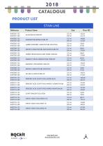
BOCiCft IM® A brand of Qujftphem www.bqckit.com www.bioquochem.com
Open the catalog to page 14
BOCkft IM® A brand of Qujftphem www.bqckit.com www.bioquochem.com
Open the catalog to page 15
BOCfc/t4 brand of Qu ^LChem [email protected] ( + 34) 985 269 292 Edificio CEEI. Parque Tecnologico Asturias. Llanera (Spain)
Open the catalog to page 16All Bioquochem catalogs and technical brochures
Archived catalogs
 PRODUCT CATALOGUE 2018
32 Pages

