CyGEL™ TECHNICAL DATA SHEET
1 /2Pages
CyGEL™ TECHNICAL DATA SHEET
1 /2Pages
Catalog excerpts

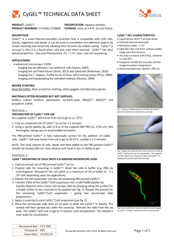
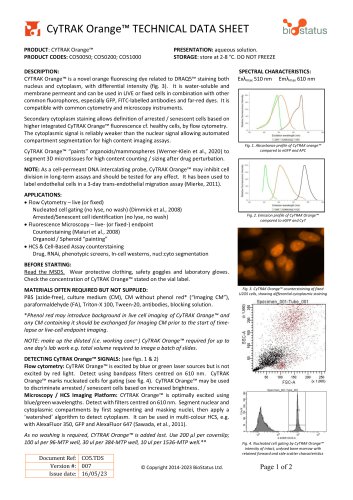
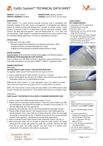
V. bidtetatus PRODUCT: CyGEL™ PRESENTATION: aqueous solution. PRODUCT NUMBERS: CY10500, CY20000 STORAGE: store at 2-8 °C. Do not freeze. DESCRIPTION: CyGEL™ is a novel thermo-reversible mountant that is compatible with LIVE cells, tissues, organisms and beads. It can be used to immobilize non-adherent objects by simple warming and conversely allowing their recovery by simple cooling. CyGEL™ is unusual in that it is a liquid when cold and a gel when warmed. CyGEL™ has ideal optical properties - low auto-fluorescence, R.I. -1.37, clear, and non-quenching. APPLICATIONS: • advanced microscopy / CLEM: imaging live de-adhered / non-adherent cells (Upton, 2007) imaging live and fixed tissues (Jecna, 2013) and spheroids (Robertson, 2010) imaging live C. elegans, fruitfly larvae (Chinta, 2012) and parasites (Price, 2009) imaging and manipulating live zebrafish embryos (Alvarez, 2009) BEFORE STARTING: Read the MSDS. Wear protective clothing, safety goggles and laboratory gloves. MATERIALS OFTEN REQUIRED BUT NOT SUPPLIED: Buffers, culture medium, plasticware, ice-bath/-pack, DRAQ5™, DRAQ7™ and propidium iodide. PROTOCOL 1: PREPARATION OF CyGEL™ FOR USE (As supplied, CyGEL™ will transit from sol to gel at ca. 23°C) 1. Cool an unopened vial of CyGEL™ on ice for 1-2 minutes. 2. Using a sterile pipette tip, add 12.8 |il of the supplied 40X PBS (i.e. 2.5% v/v). Mix thoroughly, taking care to avoid bubble formation. The PBS-primed CyGEL™ is now isotonically correct for the addition of viable cells. CyGEL™ will now transit from sol to gel at 20-21°C, usually in 1-2 minutes. NOTE: The total volume of cells, beads and dyes added to the PBS-primed CyGEL™ should not exceed 20% v/v. Over-dilution will result in loss of ability to gel. PROTOCOL 2: CyGEL™ MOUNTING OF CELLS ONTO A STANDARD MICROSCOPE SLIDE 1. Cool an unused vial of PBS-primed CyGEL™ on ice. 2. Prepare cells for mounting in CyGEL™: Wash the cells in buffer (e.g. PBS) by centrifugation. Resuspend the cell pellet in a maximum of 50 |il buffer (1 - 5 x 10⁵ cells depending upon the application). CyGEL™ KEY CHARACTERISTICS: • Liquid below 20/21°C and gel above • Infinitely thermo-reversible • Refractive index: —1.37 • Optically clear and inert, without visible range auto-fluorescence • Viscosity increases with temp., between 21 and 28°C • Excipients modify the viscosity and the sol:gel transition temperature • Recommended max. dilution: 20% v/v 3. Pipette the cell suspension into the vial containing PBS-primed CyGEL™. 4. Transfer 250ul of the CyGEL™/cell suspension into a cold P1000 pipette tip. Quickly dispense onto a clean microscope slide by streaking along the surface for a length similar to the coverslip to be applied (see fig. 1). Repeat the process for the remaining CyGEL™/cell suspension - giving two microscope slide preparations. 5. Apply a coverslip to each CyGEL™/cell suspension (see fig. 2). 6. Place the microscope slide onto an ice pack to allow the CyGEL™ to liquefy. The sample will then spread out under the coverslip. Remove the slide from the ice pack. The CyGEL™ will now re-gel as it reaches room temperature. The sample is now ready for visualization. ©Copyright BioStatus 2014-2024. All Rights Reserved
Open the catalog to page 1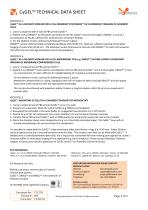
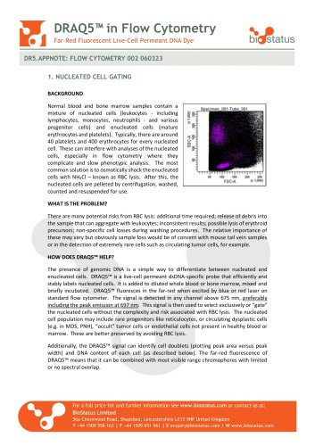
CyGEL™ TECHNICAL DATA SHEET PROTOCOL 3: CyGEL™ AS A DELIVERY MEDIUM FOR A CELL-PERMEANT DYE (DRAQ5™) IN FLUORESCENT IMAGING OF ADHERENT CELLS 1. Cool an unopened 500 ul tube of PBS-primed CyGEL™. 2. Pipette 2.05 |il DRAQ5™ (5 mM stock) and dispense into the CyGEL™ and mix thoroughly. DRAQ5™ is now at a concentration of 20 |iM, sufficient for stoichiometric chromatin binding. 3. For the addition of cells, continue by following Protocol 2 above. DRAQ5™ nuclear staining should completely equilibrate after 60-80 min. However, sufficient staining should allow imaging of nuclei after 20-30 min. The...
Open the catalog to page 2All BioStatus catalogs and technical brochures
 DRAQ9™ TECHNICAL DATA SHEET
2 Pages
 DRAQ7™ in Flow Cytometry
2 Pages
 DRAQ5™ in Flow Cytometry
2 Pages

- Solvent reagent
- Molecular biology reagent
- Research reagent
- Histology reagent
- Dye reagent kit
- Cytology reagent
- Immunoanalysis reagent kit
- Staining solution reagent
- Hematology reagent
- Flow cytometry reagent
- Immunofluorescence reagent
- DNA analysis reagent
- Blood phenotyping reagent
- Cell imaging reagent
- Hydrogel reagent