CyTRAK Orange™ TECHNICAL DATA SHEET
1 /2Pages
CyTRAK Orange™ TECHNICAL DATA SHEET
1 /2Pages
Catalog excerpts

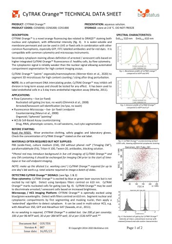
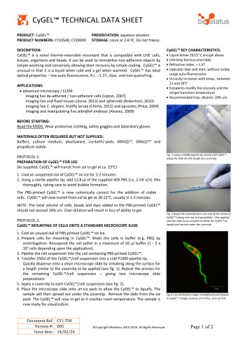
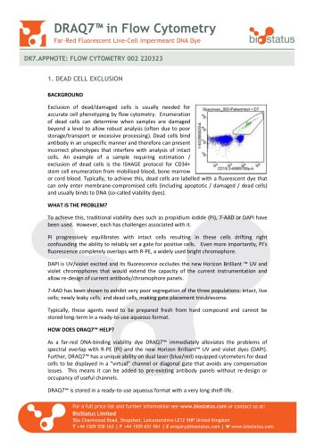
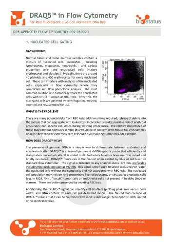
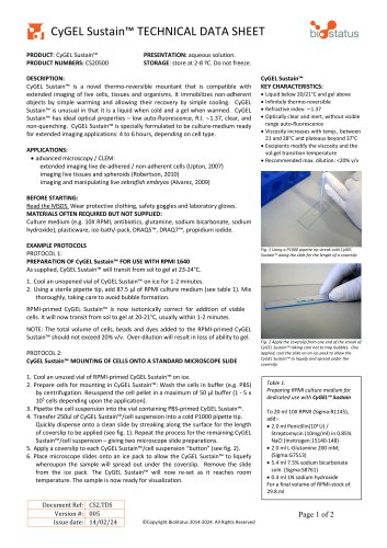
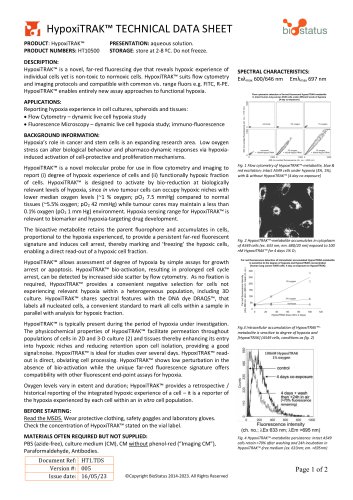
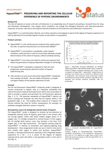
CyTRAK Orange™ TECHNICAL DATA SHEET PRODUCT: CYTRAK Orange™ PRODUCT CODES: CO50050; CO50200; CO51000 PRESENTATION: aqueous solution. STORAGE: store at 2-8 °C. DO NOT FREEZE DESCRIPTION: CYTRAK Orange™ is a novel orange fluorescing dye related to DRAQ5™ staining both nucleus and cytoplasm, with differential intensity (fig. 3). It is water-soluble and membrane permeant and can be used in LIVE or fixed cells in combination with other common fluorophores, especially GFP, FITC-labelled antibodies and far-red dyes. It is compatible with common cytometry and microscopy instruments. Secondary cytoplasm staining allows definition of arrested / senescent cells based on higher integrated CyTRAK Orange™ fluorescence cf. healthy cells, by flow cytometry. The cytoplasmic signal is reliably weaker than the nuclear signal allowing automated compartment segmentation for high content imaging assays. CyTRAK Orange™ “paints” organoids/mammospheres (Werner-Klein et al., 2020) to segment 3D microtissues for high content counting / sizing after drug perturbation. SPECTRAL CHARACTERISTICS: Exλmax 510 nm Emλmax 610 nm Fig. 1. Absorbance profile of CyTRAK orange™ compared to eGFP and APC NOTE: As a cell-permeant DNA intercalating probe, CyTRAK Orange™ may inhibit cell division in long-term assays and should be tested for any effect. It has been used to label endothelial cells in a 3-day trans-endothelial migration assay (Mierke, 2011). APPLICATIONS: • Flow Cytometry – live (or fixed) Nucleated cell gating (no lyse, no wash) (Dimmick et al., 2008) Arrested/Senescent cell identification (no lyse, no wash) • Fluorescence Microscopy – live- (or fixed-) endpoint Counterstaining (Maiuri et al., 2008) Organoid / Spheroid “painting” • HCS & Cell-Based Assay counterstaining Drug, RNAi, phenotypic screens, In-cell westerns, nucl:cyto segmentation Fig. 2. Emission profile of CyTRAK Orange™ compared to eGFP and Cy7 BEFORE STARTING: Read the MSDS. Wear protective clothing, safety goggles and laboratory gloves. Check the concentration of CyTRAK Orange™ stated on the vial label. MATERIALS OFTEN REQUIRED BUT NOT SUPPLIED: PBS (azide-free), culture medium (CM), CM without phenol red* (“Imaging CM”), paraformaldehyde (FA), Triton-X 100, Tween-20, antibodies, blocking solution. Fig. 3. CyTRAK Orange™ counterstaining of fixed U2OS cells, showing differential cytoplasmic staining *Phenol red may introduce background in live cell imaging of CyTRAK Orange™ and any CM containing it should be exchanged for Imaging CM prior to the start of timelapse or live-cell endpoint imaging. NOTE: make up the diluted (i.e. working conc n.) CyTRAK Orange™ required for up to one day’s lab work e.g. total volume required to image a batch of slides. DETECTING CyTRAK Orange™ SIGNALS: (see figs. 1 & 2) Flow cytometry: CyTRAK Orange™ is excited by blue or green laser sources but is not excited by red light. Detect using bandpass filters centred on 610 nm. CyTRAK Orange™ marks nucleated cells for gating (see fig. 4). CyTRAK Orange™ may be used to discriminate arrested / senescent cells based on increased brightness. Microscopy / HCS Imaging Platform: CYTRAK Orange™ is optimally excited using blue/green wavelengths. Detect with filters centred on 610 nm. Segment nuclear and cytoplasmic compartments by first segmenting and masking nuclei, then apply a ‘watershed’ algorithm to detect cytoplasm. It can be used in multi-colour HCS, e.g. with AlexaFluor 350, GFP and AlexaFluor 647 (Sawada, et al., 2011). As no washing is required, CYTRAK Orange™ is added last. Use 200 µl per coverslip; 100 ul per 96-MTP well, 30 ul per 384-MTP well, 10 ul per 1536-MTP well.** Document Ref: CO5.TDS Version #: 007 Issue date: 16/05/23 Fig. 4. Nucleated cell gating by CyTRAK Orange™ intensity of intact, unlysed bone marr
Open the catalog to page 1
>dft biSstatus EXAMPLE PROTOCOLS PROTOCOL 1: NUCLEATED AND SENESCENT/ARRESTED CELL GATING BY FLOW OR IMAGING CYTOMETRY 1. Prepare cells for staining with CYTRAK Orange™: resuspend cells in appropriate buffer (PBS) at a concentration of <4 x 10⁵ / ml in a test tube. For adherent cells estimate the number of cells based on confluence level or tissue section size. 2. Add CYTRAK Orange™ at 5-10 pM, final concentration. This will be an overlay for adherent cells / tissue sections, added to the well directly or in fresh medium following a wash step. 3. Gently mix, then incubate for 15-30 minutes at...
Open the catalog to page 2All BioStatus catalogs and technical brochures
 CyGEL™ TECHNICAL DATA SHEET
2 Pages
 DRAQ9™ TECHNICAL DATA SHEET
2 Pages
 DRAQ7™ in Flow Cytometry
2 Pages
 DRAQ5™ in Flow Cytometry
2 Pages

- Solvent reagent
- Molecular biology reagent
- Research reagent
- Histology reagent
- Dye reagent kit
- Cytology reagent
- Immunoanalysis reagent kit
- Staining solution reagent
- Hematology reagent
- Flow cytometry reagent
- Immunofluorescence reagent
- DNA analysis reagent
- Blood phenotyping reagent
- Cell imaging reagent
- Hydrogel reagent