DRAQ5™ in Flow Cytometry
1 /2Pages
DRAQ5™ in Flow Cytometry
1 /2Pages
Catalog excerpts

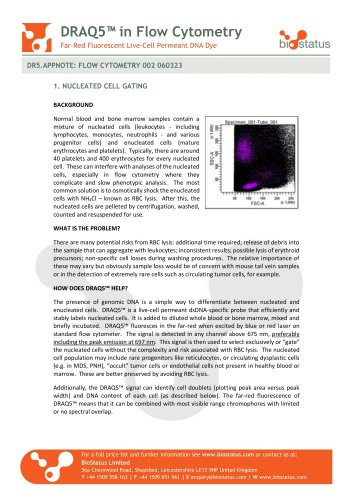
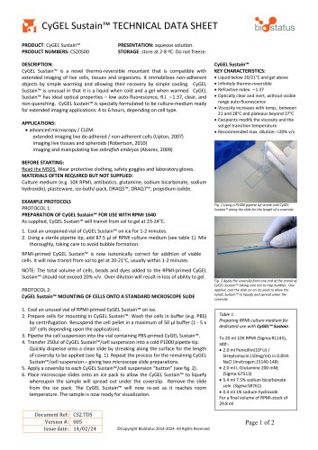
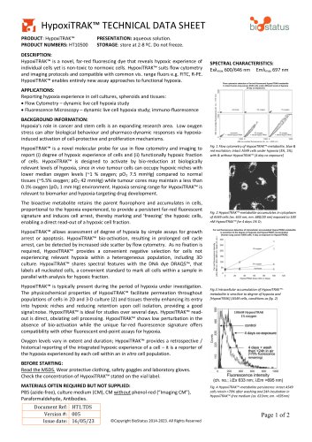
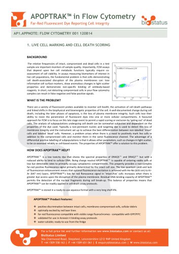
DRAQ5™ in Flow Cytometry Far-Red Fluorescent Live-Cell Permeant DNA Dye DR5.APPNOTE: FLOW CYTOMETRY 002 060323 1. NUCLEATED CELL GATING BACKGROUND Normal blood and bone marrow samples contain a mixture of nucleated cells (leukocytes - including lymphocytes, monocytes, neutrophils - and various progenitor cells) and enucleated cells (mature erythrocytes and platelets). Typically, there are around 40 platelets and 400 erythrocytes for every nucleated cell. These can interfere with analyses of the nucleated cells, especially in flow cytometry where they complicate and slow phenotypic analysis. The most common solution is to osmotically shock the enucleated cells with NH4Cl – known as RBC lysis. After this, the nucleated cells are pelleted by centrifugation, washed, counted and resuspended for use. WHAT IS THE PROBLEM? There are many potential risks from RBC lysis: additional time required; release of debris into the sample that can aggregate with leukocytes; inconsistent results; possible lysis of erythroid precursors; non-specific cell losses during washing procedures. The relative importance of these may vary but obviously sample loss would be of concern with mouse tail vein samples or in the detection of extremely rare cells such as circulating tumor cells, for example. HOW DOES DRAQ5™ HELP? The presence of genomic DNA is a simple way to differentiate between nucleated and enucleated cells. DRAQ5™ is a live-cell permeant dsDNA-specific probe that efficiently and stably labels nucleated cells. It is added to diluted whole blood or bone marrow, mixed and briefly incubated. DRAQ5™ fluoresces in the far-red when excited by blue or red laser on standard flow cytometer. The signal is detected in any channel above 675 nm, preferably including the peak emission at 697 nm. This signal is then used to select exclusively or “gate” the nucleated cells without the complexity and risk associated with RBC lysis. The nucleated cell population may include rare progenitors like reticulocytes, or circulating dysplastic cells (e.g. in MDS, PNH), “occult” tumor cells or endothelial cells not present in healthy blood or marrow. These are better preserved by avoiding RBC lysis. Additionally, the DRAQ5™ signal can identify cell doublets (plotting peak area versus peak width) and DNA content of each cell (as described below). The far-red fluorescence of DRAQ5™ means that it can be combined with most visible range chromophores with limited or no spectral overlap. For a full price list and further information see www.biostatus.com or contact us at: BioStatus Limited 56a Charnwood Road, Shepshed, Leicestershire LE12 9NP United Kingdom T +44 1509 558 163 | F +44 1509 651 061 | E [email protected] |
Open the catalog to page 1
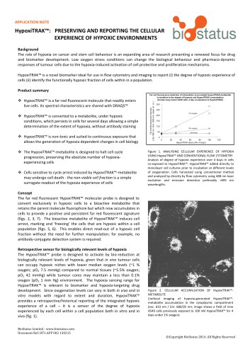
2. DNA CONTENT (OR CELL CYCLE) ANALYSIS BACKGROUND The cell cycle describes a series of events that occur when a cell divides and replicates into two daughter cells. Cell cycle regulation depends upon processes that are pivotal to cell survival. During the cell cycle the quantity of DNA increases from 2N (G1 phase) to 4N (G2 phase) via the S (synthesis) phase. Perturbation of the cell cycle can lead, for example, to cell cycle arrest or uncontrolled cell division, as in cancer, while cells with DNA below 2N are typically in apoptosis. The quantity of DNA in each nucleated cell in a population...
Open the catalog to page 2All BioStatus catalogs and technical brochures
 CyGEL™ TECHNICAL DATA SHEET
2 Pages
 DRAQ9™ TECHNICAL DATA SHEET
2 Pages
 DRAQ7™ in Flow Cytometry
2 Pages

- Solvent reagent
- Molecular biology reagent
- Research reagent
- Histology reagent
- Cytology reagent
- Dye reagent kit
- Immunoanalysis reagent kit
- Staining solution reagent
- Hematology reagent
- Flow cytometry reagent
- Immunofluorescence reagent
- DNA analysis reagent
- Blood phenotyping reagent
- Cell imaging reagent
- Hydrogel reagent