DRAQ9™ in Cell Painting / Mosaic
1 /1Page

DRAQ9™ in Cell Painting / Mosaic
1 /1Page
Catalog excerpts

DRAQ9™ in Cell Painting / Mosaic Far-Red Fluorescent Live-Cell Tracking & Painting Dye DR9.APPNOTE: PAINTING 001 070617 1. PHENOTYPIC SCREENING BACKGROUND High content phenotypic screening has accelerated drug discovery programmes without target knowledge. This is typically achieved using a cell type with appropriate disease phenotype(s) from which a readout can be obtained. In oncology the goal may be cell killing, catastrophic metabolic event or cell arrest. The change could, however, be at one of many possible loci in the cellular machinery and outisde oncology could be illicited more subtly. Fluorescence-based imaging necessitates use of either transformed cells expressing fluorescent proteins in mechanisms, compartments or sentinel pathways and/or the use of fluorescent probes in the case of cells without such genetically-engineered elements or where additional subcellular demarcation is required. Image analysis software can thereafter determine cells with relevant changes that suggest further investigation of the implicated molecular entity to determine the nature of the interaction, perhaps including identification of the target(s). WHAT IS THE PROBLEM? Working with primary cells a fluorescent-reporter is unavailable. It may then be necessary to use a battalion of target–specific fluorescent probes in the attempt to uncover a phenotypic change under the influence of the compound library being screened. At scale this can be limiting since the useful readout may elude the options available within the confines of the fluorescence spectrum. Similarly, such a multi-probe approach will be costly. Moreover, even with transformed cells containing a GFP-reporter, the reporter may provide a useful readout. An alternative approach is use of a fluorescent probe which “decorates” the cell in a predictable way not relying upon specific targeting but rather uses a training set for the different cellular states that are exemplars. In this way it is possible to interrogate cells for a phenotypic change imposed by the chemical entity and/or its dose using a pattern change within the cytoplasm. It would be helpful if this could be achieved on live cells prior to fixation or post-fixation depending on the assay workflow, with some defined gross pattern for cellular morphology such as the cytoplasmic boundary or the nucleus. The probe should permit a wide range of other fluorescent probes to be combined with it e.g. mitochondrial probe or nuclear counterstain. HOW DOES DRAQ9™ HELP? The far-red, live cell permeant cytoplasmic probe DRAQ9™ provides staining of cells, either live or fixed, that can be the basis of a non-a priori phenotypic screening method since it labels a range of features in the cytoplasm of cells. Helpfully, it is “nuclear-dark” to leave a well-established morphological feature for a nuclear counterstain, if required. The emission spectrum means it is highly compatible with visible-range and UV-excited chromophores. Practically, DRAQ9™ is provided in an aqueous, ready-to-use solution and can be admixed with formaldehyde fixative for a single-step fix and stain procedure. far-red fluorescing cell permeant cytoplasmic probe labels peri-nuclear structures e.g. Golgi, ER, IVTS optimally excited by red laser lines, no UV requirement compatible with UV-excited and visible range chromophores water-soluble; ready-to-use from the fridge For a full price list and further information see www.biostatus.com or contact us at: BioStatus Limited 56a Charnwood Road, Shepshed, Leicestershire LE12 9NP United Kingdom T +44 1509 558 163 | F +44 1509 651 061 | E enquiry@biostatu
Open the catalog to page 1All BioStatus catalogs and technical brochures
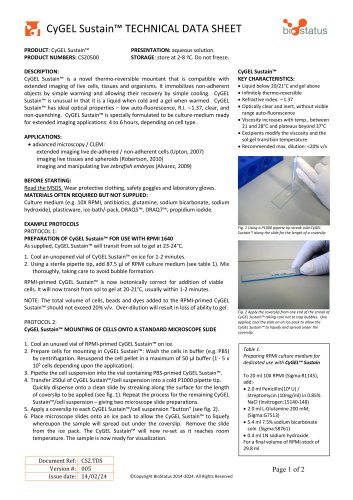
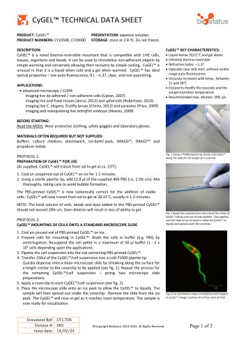 CyGEL™ TECHNICAL DATA SHEET
2 Pages
 DRAQ9™ TECHNICAL DATA SHEET
2 Pages
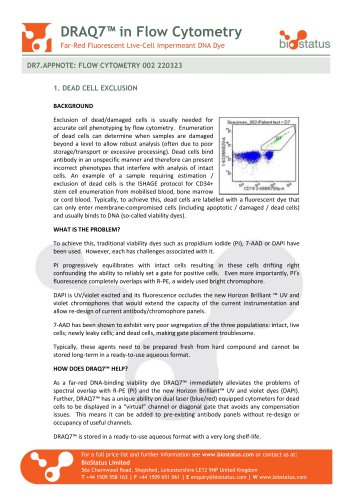 DRAQ7™ in Flow Cytometry
2 Pages
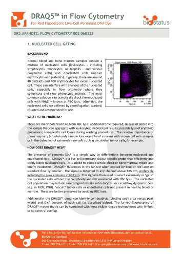 DRAQ5™ in Flow Cytometry
2 Pages
- Solvent reagent
- Molecular biology reagent
- Research reagent
- Histology reagent
- Dye reagent kit
- Cytology reagent
- Immunoanalysis reagent kit
- Staining solution reagent
- Hematology reagent
- Flow cytometry reagent
- Immunofluorescence reagent
- DNA analysis reagent
- Blood phenotyping reagent
- Cell imaging reagent
- Hydrogel reagent