HypoxiTRAK™ TECHNICAL DATA SHEET
1 /2Pages
HypoxiTRAK™ TECHNICAL DATA SHEET
1 /2Pages
Catalog excerpts

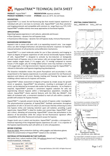
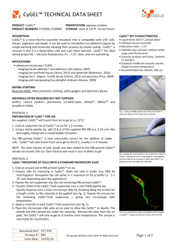
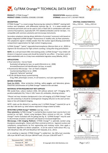
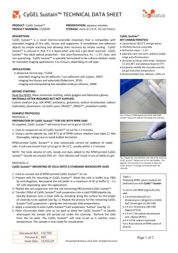
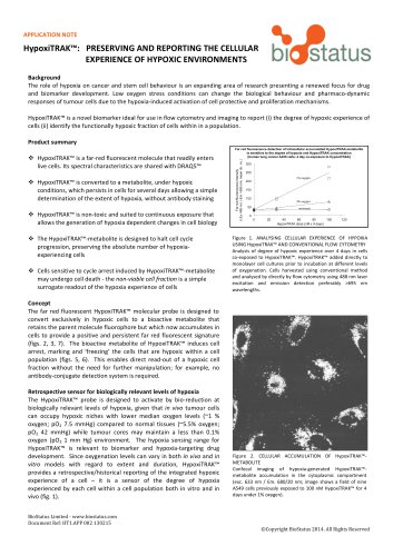
HypoxiTRAK™ TECHNICAL DATA SHEET PRODUCT: HypoxiTRAK™ PRODUCT NUMBERS: HT10500 PRESENTATION: aqueous solution. STORAGE: store at 2-8 ºC. Do not freeze. DESCRIPTION: HypoxiTRAK™ is a novel, far-red fluorescing dye that reveals hypoxic experience of individual cells yet is non-toxic to normoxic cells. HypoxiTRAK™ suits flow cytometry and imaging protocols and compatible with common vis. range fluors e.g. FITC, R-PE. HypoxiTRAK™ enables entirely new assay approaches to functional hypoxia. APPLICATIONS: Reporting hypoxia experience in cell cultures, spheroids and tissues: • Flow Cytometry – dynamic live cell hypoxia study • Fluorescence Microscopy – dynamic live cell hypoxia study; immuno-fluorescence BACKGROUND INFORMATION: Hypoxia’s role in cancer and stem cells is an expanding research area. Low oxygen stress can alter biological behaviour and pharmaco-dynamic responses via hypoxiainduced activation of cell-protective and proliferation mechanisms. HypoxiTRAK™ is a novel molecular probe for use in flow cytometry and imaging to report (i) degree of hypoxic experience of cells and (ii) functionally hypoxic fraction of cells. HypoxiTRAK™ is designed to activate by bio-reduction at biologically relevant levels of hypoxia, since in vivo tumour cells can occupy hypoxic niches with lower median oxygen levels (~1 % oxygen; pO 2 7.5 mmHg) compared to normal tissues (~5.5% oxygen; pO2 42 mmHg) while tumour cores may maintain a less than 0.1% oxygen (pO2 1 mm Hg) environment. Hypoxia sensing range for HypoxiTRAK™ is relevant to biomarker and hypoxia-targeting drug development. The bioactive metabolite retains the parent fluorophore and accumulates in cells, proportional to the hypoxia experienced, to provide a persistent far-red fluorescent signature and induces cell arrest, thereby marking and ‘freezing’ the hypoxic cells, enabling a direct read-out of a hypoxic cell fraction. SPECTRAL CHARACTERISTICS: Exλmax 600/646 nm Emλmax 697 nm Flow cytometric detection of far-red fluorescent HypoxiTRAK-metabolite in intact human lung cancer A549 cells under different levels of hypoxia [4 day co-exposure] Fig. 1 Flow cytometry of HypoxiTRAK™-metabolite, blue & red excitation; intact A549 cells under hypoxia (3%, 1%), with & without HypoxiTRAK™ [4 day co-exposure] Fig. 2 HypoxiTRAK™-metabolite accumulates in cytoplasm of A549 cells (ex. 633 nm; em. 680/20 nm) exposed to 100 nM HypoxiTRAK™ for 4 days 1% O 2 HypoxiTRAK™ allows assessment of degree of hypoxia by simple assays for growth arrest or apoptosis. HypoxiTRAK™ bio-activation, resulting in prolonged cell cycle arrest, can be detected by increased side scatter by flow cytometry. As no fixation is required, HypoxiTRAK™ provides a convenient negative selection for cells not experiencing relevant hypoxia within a heterogeneous population, including 3D culture. HypoxiTRAK™ shares spectral features with the DNA dye DRAQ5™, that labels all nucleated cells, a convenient standard to mark all cells within a sample in parallel with analysis for hypoxic fraction. HypoxiTRAK™ is typically present during the period of hypoxia under investigation. The physicochemical properties of HypoxiTRAK™ facilitate permeation throughout populations of cells in 2D and 3-D culture (2) and tissues thereby enhancing its entry into hypoxic niches and reducing retention upon cell isolation, providing a good signal:noise. HypoxiTRAK™ is ideal for studies over several days. HypoxiTRAK™ readout is direct, obviating cell processing. HypoxiTRAK™ shows low perturbation in the absence of bio-activation while the unique far-red fluorescence signature offers compatibility with other fluorescent end-point assays for hypoxia. Fig.3 Intracellular accumulation of HypoxiTRAK™metabolite is sensitive to degree of hypoxia and [HypoxiTRAK] (A549 cells, conditions as fig. 2) Oxygen levels vary in extent and duration; HypoxiTRAK™ provides a retrospective / historical reporting of the integrated hypoxic experience of a cell – it is a reporter of the hypoxia experienced by each cell within an in vitro cell population. BEFORE STARTING: Read the MSDS. Wear protective clothing, safety goggles and laboratory gloves. Check the concentration of HypoxiTRAK™ stated on the vial label. MATERIALS OFTEN REQUIRED BUT NOT SUPPLIED: PBS (azide-free), culture medium (CM), CM without phenol-red (“Imaging CM”), Paraformaldehyde, Antibodies. Document Ref: HT1.TDS Version #: 005 Issue date: 16/05/23 Fig. 4 HypoxiTRAK™-metabolite persistence: intact A549 cells retain >70% after washing and 24h incubation in HypoxiTRAK™-free medium (ex. 633nm; em
Open the catalog to page 1
FOR FURTHER INFORMATION PLEASE CONTACT: BioStatus Limited Website: www.biostatus.com Technical Support: [email protected] Re-ordering: [email protected] BioStatus Limited 56A Charnwood Road, Shepshed, Leicestershire LE12 9NP UK Company registered in England and Wales. No. 3079239 HypoxiTRAK™ TECHNICAL DATA SHEET **NOTE: retained HypoxiTRAK™-metabolite inhibits cell division - marking and preserving cells that have experienced hypoxic conditions. DETECTING HypoxiTRAK™ SIGNALS: (see Fig. 1) Flow cytometry: HypoxiTRAK™-metabolite can be excited by 488 or 635 nm wavelength light. Detect in...
Open the catalog to page 2All BioStatus catalogs and technical brochures
 CyGEL™ TECHNICAL DATA SHEET
2 Pages
 DRAQ9™ TECHNICAL DATA SHEET
2 Pages
 DRAQ7™ in Flow Cytometry
2 Pages
 DRAQ5™ in Flow Cytometry
2 Pages

- Solvent reagent
- Molecular biology reagent
- Research reagent
- Histology reagent
- Cytology reagent
- Dye reagent kit
- Immunoanalysis reagent kit
- Staining solution reagent
- Hematology reagent
- Flow cytometry reagent
- Immunofluorescence reagent
- DNA analysis reagent
- Blood phenotyping reagent
- Cell imaging reagent
- Hydrogel reagent