- Catalogs
- Boeckeler Instruments, Inc.
- Matsci-B1
Matsci-B1

Matsci-B1
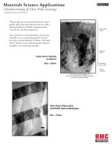
This bulletin discusses the application of ultramicrotomy in the preparation of thin film coatings for materials science applications. The technique involves using a diamond knife on an RMC ultramicrotome to cut ultra-thin sections for electron microscopy analysis.
Specifications
The sections are cut at a speed of 0.1mm/sec with a thickness of 40nm, facilitated by the Power Drive© feature of the ultramicrotomes. This allows for the sectioning of extremely hard materials.
Applications
Ultramicrotomy is used to cross-section hard semiconductor, dielectric, and metal coatings in the electronics and optics industries. It enables the study of structure/property relationships in thin films using Transmission Electron Microscopy and qualitative elemental analysis by EDS.
Sample Preparation
Samples are prepared from micro-chips approximately 1000 microns in diameter, cleaned, and embedded in Spurrs epoxy resin before sectioning.
Examples
- Cubic Boron Nitride on Silicon (Bar = 25nm)
- PS E-beam Deposited TiO2/SiO2 Optical Multilayer (Bar = 25nm)
- E-beam Evaporated Deposition of Fluoride Compound on Glass Substrate (Bar = 200nm)
- ZnS/SiO2 Multilayer Optical Coating (Bar = 50nm)
Contact Information
Boeckeler Instruments, Inc.
4650 South Butterfield Drive, Tucson, AZ 85714, U.S.A.
Phone: (520) 745-0001
Fax: (520) 745-0004
Email: [email protected]
Website: www.rmcboeckeler.com
© 2015 Boeckeler Instruments, Inc. All specifications subject to change without notice.
Catalog excerpts
These images are cross-sectional electron micrographs taken from ultra-thin sections cut with a diamond knife on an RMC ultramicrotome. (see MT-XL and MT-X datasheets) The materials are extremely hard so sections are typically cut at a sectioning speed of 0.1mm/ sec with a section thickness of 40nm. The Power Drive© feature of our ultramicrotomes makes it possible to cut such hard materials. Cubic Boron Nitride on Silicon Bar = 25nm PS E-beam Deposited Ti02/SiO2 Optical Multilayer Bar = 25nm
Open the catalog to page 1
Ultra-thin sectioning with an ultramicrotome is frequently used to cross-section hard semiconductor, dialectric and metal coatings used in the electronics and optics industry. This method provides a critical capability to study the structure/property relationships in thin films using Transmission Electron Microscopy and qualitative elemental analysis by EDS. Samples are typically prepared from micro-chips in the order of 1000 microns in diameter, cleaned and embedded in Spurrs epoxy resin prior to sectioning on the ultramicrotome. E-beam Evaporated Deposition of Fluoride Compound on Glass Substrate...
Open the catalog to page 2All Boeckeler Instruments, Inc. catalogs and technical brochures
 Ultramicrotomy PTXL
3 Pages
 PTPCZ
PTPCZ5 Pages
 ASH2
ASH22 Pages
 Array Tomography ATUMtome
4 Pages
 Cryo Ultramicrotomy LN Ultra
6 Pages
 Jeol JFD-V
2 Pages
 HPM 010 3-fold
2 Pages
 HPM 010
2 Pages
 GKM-2 3-fold
2 Pages
 GKM-2
GKM-22 Pages
 FS-8500
2 Pages
 EMP-5160
2 Pages
 Diamond Knives
1 Page
 CRT-900 Cryo Accessory
2 Pages
 CR-X Ultracut
2 Pages
 CR-X
CR-X2 Pages
 ASH-100
2 Pages
 AFM-990 Cryo
2 Pages
 AFM-990
2 Pages
 60617-SR
2 Pages
 RMC Boeckeler Line Card 0517
2 Pages
 MT-990
2 Pages
 MR2, MR3
2 Pages
 Matsci-B2
2 Pages
 LN-Ultra Cryo System
2 Pages
 HPM Live Mu
2 Pages
 QG-3100
2 Pages
 sample prep solutions
12 Pages

- Sample preparation system
- Automatic sample processor
- Laboratory sample preparation system
- Benchtop sample preparation system
- Tissue processor
- Slide stainer
- Rotary microtome
- Embedding system
- Research sample preparation system
- Motorized microtome
- Automatic microtome
- Rotary microtome cryostat
- Manual microtome
- Programmable microtome
- Automatic microtome cryostat
- Motorized microtome cryostat
- Electron microscopy sample preparation system
- Automatic ultramicrotome
- Rotary ultramicrotome