- Products
- Catalogs
- News & Trends
- Exhibitions
AUTOMATIC BRAIN METASTASES PLANNING Clinical White Paper
AUTOMATIC BRAIN METASTASES PLANNING Clinical White Paper
Radiosurgery is a primary treatment for patients with multiple brain metastases, focusing on cumulative tumor volume rather than the number of metastases. Brainlab's Automatic Brain Metastases Planning targets up to ten metastases simultaneously, optimizing treatment quality and efficiency.
The Assuta Medical Center in Israel has implemented this technology, reviewing ten early cases. Patients were over 50 years old with 2 to 19 metastases, primarily from lung cancer. Lesions were identified using MRI and CT imaging.
Data is imported into the Elements platform, where tumor volumes are contoured. Critical organs are automatically segmented. Treatment templates are predefined, considering tumor volume, prescription doses, and gantry angles. The isocenter is determined by the average position of target centers, and arcs are optimized to minimize exposure to healthy tissue.
Arc weights are optimized for conformity using the inverse Paddick conformity index. The treatment plan is optimized in under two minutes. Novalis frameless radiosurgery technology is used, with a thermoplastic mask ensuring patient stability.
ExacTrac provides image-guided positioning, allowing for intra-fraction monitoring. It ensures sub-millimeter accuracy by correcting for patient movement and table motion.
Automatic Brain Metastases Planning enhances radiosurgery by improving treatment efficiency and quality, supported by advanced imaging and positioning technologies.
Catalog excerpts

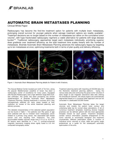
AUTOMATIC BRAIN METASTASES PLANNING Clinical White Paper Radiosurgery has become the first-line treatment option for patients with multiple brain metastases, prolonging overall survival for younger patients when salvage treatment options are readily available1. Treatment decisions are no longer based on the number of metastases but rather on the cumulative tumor volume2, with hypo-fractionated radiosurgery regimens a viable alternative for patients with large disease burden3-6. Traditional radiosurgery approaches target each metastasis individually; prioritizing superior dose gradients over treatment efficiency as the total treatment time scales linearly with the number of metastases. Brainlab Automatic Brain Metastases Planning advances the radiosurgery legacy by targeting up to ten metastases at once; optimizing treatments both in terms of plan quality and delivery efficiency. Figure 1: Automatic Brain Metastases Planning details for Patient 4 with 9 lesions. The Assuta Medical Center located just north of Tel Aviv, along the popular Mediterranean coastline of Israel, has been a radiosurgery center of excellence for many years. Equipped with Novalis Radiosurgery and a high definition large field multileaf collimator, this center has been pioneering the clinical implementation of Brainlab Automatic Brain Metastases Treatment Planning. Starting treatments in summer 2014, they retrospectively selected ten early cases treated at their institution for review of the entire treatment planning and delivery process. The detailed characteristics of the selected patient population can be found in Table 1. Ten patients over fifty years of age were selected with two to nineteen brain metastases; seven related to a primary lung cancer, two associated with breast cancer and one resulting from melanoma. The total accumulated treated volumes range from 0.15 cc to 19.78 cc. All lesions were identified on 1 mm T1 contrast-enhanced magnetic resonance imaging slices rigidly fused to 0.6 mm computed tomography slices. Treatment planning starts with importing all DICOM data into the Elements treatment planning platform, fusing the various data sets and contouring the gross tumor volumes of each target on the magnetic resonance data sets utilizing the Smartbrush Element. All critical organs are automatically segmented based on a universal atlas approach and can be consulted in a dedicated DICOM Viewer. Automatic Brain Metastases Planning takes the target volumes and models the patient outer contour and underlying table top for correct dose calculations. A list of numerous treatment templates matching diverse hospital practices can be defined upfront with the most appropriate template chosen for each patient. Treatment templates contain various information: possible enlargement of the tumor volumes to encompass biological and treatment uncertainties, different prescription doses in function of individual tumor volumes or sizes, table and gantry angle preferences as a guide for the subsequent automatic optimization of the arcs.
Open the catalog to page 1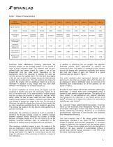
Table 1: Patient Characteristics Automatic Brain Metastases Planning determines the isocenter position as the average position of the centers of mass of each individual target. The software uses the predefined table and gantry angles to create a set of two independent arcs per table angle. Depending on the hemisphere where the isocenter is located, the arcs are mirrored around the sagittal plane. The start and stop angles of the arc are first set to the template values and automatically modified during optimization. The leaf positions are shaped to conform the targets, with an additional margin...
Open the catalog to page 2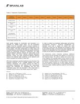
Table 2: Treatment Characteristics Inverse Paddick Conformity Index High quality imaging for localization and simulation is a necessity for reliable image fusion. For the localization images, ExacTrac accredits superior contrast resolution in optimal exposure conditions to the unique configuration of the X-Ray system. The fixed configuration of X-Ray tubes and flat-panel detectors eliminates any potential spatial uncertainty caused by mechanical movement. Furthermore, the large source-to-detector distance reduces the solid angle of the radiation beam and hence reduces potential geometric distortion....
Open the catalog to page 3All Brainlab catalogs and technical brochures
 CRANIAL ACCESSORIES
13 Pages
 Buzz In-Wall
6 Pages
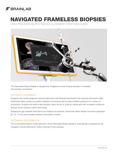 NAVIGATED FRAMELESS BIOPSIES
2 Pages


Archived catalogs
 Brochure Novalis
2 Pages
 VoyantMark
1 Page
 iPlan Flow
2 Pages
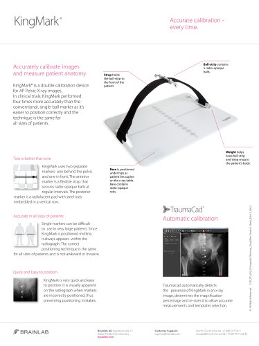 KingMark
1 Page
 LIVE IMAGING
2 Pages
 Radiolucent Skull Pins
1 Page
 Product Catalog Neurosurgery
9 Pages
 Hip Navigation
23 Pages
 Elements
14 Pages
 Quentry
12 Pages
 Integrated MR
1 Page
 Cranial Navigation
2 Pages
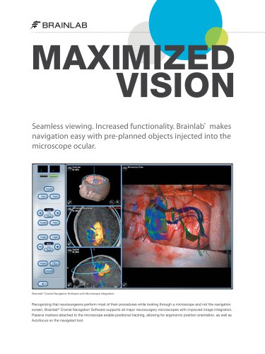 Microscope Integration
2 Pages
 KNEE 3
15 Pages
 Curve
Curve18 Pages
 Flyer Hip Express
2 Pages
 AIRO
AIRO10 Pages
 Flyer Knee Express
2 Pages
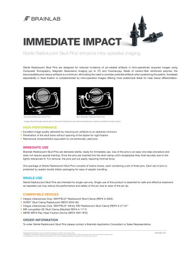
 IMMEDIATE IMPACT
1 Page
 Brochure Spine Navigation
19 Pages
 Vero sBrt unleashed
12 Pages
 Buzz
Buzz12 Pages
 iPlan Cranial
2 Pages
 iPlan HybridArc
2 Pages
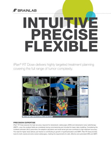 iPlan RT Dose
2 Pages
 iPlan Monte Carlo
2 Pages
 iPlan RT Adaptive
2 Pages
 iPlan
iPlan5 Pages


- Analysis software
- Visualization software
- Radiology software
- Tablet computer software
- Tablet PC software
- Control software
- Windows software
- Diagnostic software
- Planning software
- Cloud-based software
- Hospital software
- Automated software
- Treatment software
- Traceability software
- Software module
- Surgery software
- Training software
- Import software
- Sharing software
- CT software