- Catalogs
- Bruker Daltonics Inc.
- Understanding Primary Metastasis of Ovarian Cancer via Imaging Mass Spectrometry of a Novel 3D Tissue Explant and Cellular Coculture
Understanding Primary Metastasis of Ovarian Cancer via Imaging Mass Spectrometry of a Novel 3D Tissue Explant and Cellular Coculture
1 /8Pages

Understanding Primary Metastasis of Ovarian Cancer via Imaging Mass Spectrometry of a Novel 3D Tissue Explant and Cellular Coculture
1 /8Pages
Catalog excerpts
Understanding Primary Metastasis of Ovarian Cancer via Imaging Mass Spectrometry of a Novel 3D Tissue Explant and Cellular Coculture Cellular and molecular signaling have important roles in the development and progression of cancer. However, as this communication is often dynamic and environment-dependent, it is difficult to characterize using traditional analytical techniques. A specific example of this challenge is to understand the primary metastasis of high grade serous ovarian cancer (HGSOC). It has been suggested that HGSOC first develops in the fallopian tube, and then transformed carcinogenic cells migrate to Authors: Dr. Laura Sanchez, Katherine Zink; University of Illinois at Chicago, Chicago, IL, USA. the ovary to form fallopian-tubeepithelium-derived (FTE-derived) HGSOC. Keywords: MALDI-TOF, autoflex, MALDI Imaging, ovarian cancer
Open the catalog to page 1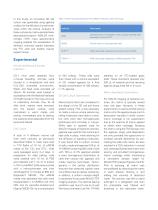
In this study, an innovative 3D cell culture was generated using agarose to allow for the diffusion of small molecules within the culture. Analysis of these cultures by matrix-assisted laser desorption/ionization (MALDI) timeof-flight (TOF) mass spectrometry imaging enabled the visualization of different chemical signals between the FTE cells and healthy ovarian explant tissue. Table 1: Names and descriptions of the 4 different cell lines used in this study. Cell Line Spontaneously immortalized murine oviductal epithelial cells (equivalent of human FTE) Murine ovarian surface epithelial cells...
Open the catalog to page 2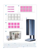
Figure 1: Overview of workflow for MALDI Imaging. A Ovaries from 16-18 day old female mice were extracted and maintained in warm media. B Ovaries are separated from bursa and halved immediately prior to plating. Explants are placed in the center of half of the wells of the 8-well chamber mounted to an ITO-coated glass slide. C 300 µL of various cell cultures prepared in 1% agarose are plated in each well. Each cell type was plated with an ovarian explant and as a pure cell culture. The entire slide was covered with a lid and incubated for 4 days at 5% CO2 and 37°C. D The 8-well chamber is removed....
Open the catalog to page 3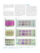
The solution was immediately filtered through a fritted glass filter, and the crystals were collected and dried under air and then under a high vacuum. MALDI-TOF Imaging Samples were first scanned at 1200 dpi to integrate an image for data acquisition. Then, samples were analyzed using a Bruker autoflex speed LRF mass spectrometer over m/z 100-2000 Da. The instrument was calibrated manually using red phosphorus and operated in positive mode, with laser power set to 40%, laser width set to 2 (small) and reflector gain set to 2.0x. At each sample location, 500 laser shots were fired at 2000 Hz...
Open the catalog to page 4
A subsequent experiment was then performed to determine if these 44 m/z signals were unique to tumori-genic FTE-derived cells. Eight separate conditions were prepared on one slide: (1) agarose and media only (control), (2) an ovarian explant alone, (3) MOE SCRshRNA cells, (4) an ovarian and MOE SCRshRNA cell coculture, (5) MOE PTENshRNA cells, (6) an ovarian explant and MOE PTENshRNA cells coculture, (7) MOSE cells, and (8) an ovarian explant and MOSE cell coculture. The ovarian explant and MOE SCRshRNA cell coculture served as a control to determine if the observed signals were also elicited...
Open the catalog to page 5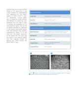
However, due to the mixed coculture design of this experiment, it was unclear whether the norepinephrine was originating from the ovarian explant or MOE PTENshRNA cells. An experiment utilizing plastic tabs to divide the 8-well chamber was designed to embed the ovary separately from the cells in order to visualize the localization of the norepinephrine signal. Nine of the 33 signals identified as significant in the original experimental design were also detected here, including norepinephrine (Figure 5). It was clear from the spatial distribution of the signals that the ovary was the source of...
Open the catalog to page 6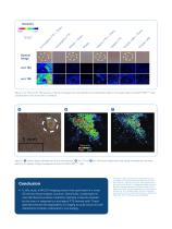
Figure 4: m/z 104 and m/z 136 were two of the 33 m/z signals that were identified to be significantly higher in the ovarian explant and MOE PTENshRNA cells coculture than in any of the other 7 conditions. Figure 5: A Optical image of divided well of an 8-well chamber; B m/z 112 and C m/z 144 are two signals that were clearly originated from the ovary explant and migrated through the agarose towards the MOE PTENshRNA cells. Conclusion • In this study, a MALDI Imaging protocol was optimized for a novel 3D cell and tissue explant coculture. Specifically, norepinephrine was identified as putative...
Open the catalog to page 7
Learn More You are looking for further Information? Check out the link or scan the QR code for more details. Bruker Daltonik GmbH Bremen · Germany Phone +49 (0)421-2205-0 [email protected] – www.bruker.com to change specifications without notice. © Bruker Daltonics 04-2019, MSI-09, 1868198 For Research Use Only. Not for Use in Clinical Diagnostic Procedures. Bruker Daltonics is continually improving its products and reserves the right
Open the catalog to page 8All Bruker Daltonics Inc. catalogs and technical brochures
 Life Science Mass Spectrometry
20 Pages
 MASS SPECTROMETRY
20 Pages
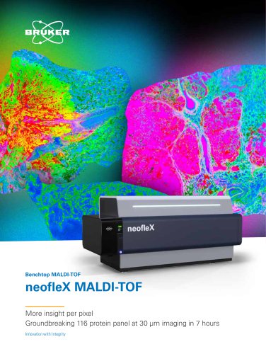 neofleX™
12 Pages
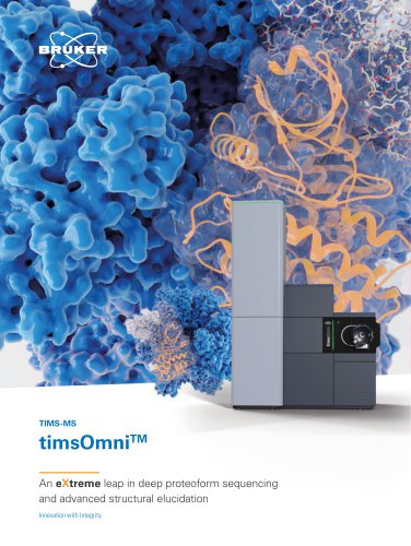 timsOmniTM
20 Pages
Archived catalogs
 micrOTOF-Q III
6 Pages
 Toxtyper
6 Pages
 Radiation Backpack Sentry
4 Pages
 impact HD
12 Pages
 Product Overview
20 Pages
 The new autoflex speed
10 Pages
 solarix XR
12 Pages
 SIGIS II
2 Pages
 VeroTect
4 Pages
 µRAID
µRAID4 Pages
 SVG 2 and Probes
6 Pages
 MM2
MM26 Pages
 RAID M100
6 Pages
 RAID S2
6 Pages
 RAID XP
4 Pages
 RAID AFM
4 Pages
 pTD
pTD2 Pages
 DE-tector
6 Pages
 ImagePrep
4 Pages
 micrOTOF II
8 Pages
 impact II
12 Pages
 EVOQ
EVOQ6 Pages
 maxis II
12 Pages
 ProteinScape
8 Pages
 Bruker ToxScreener
6 Pages
 MALDI Biotyper CA System
12 Pages
 MALDI Biotyper Clinical IVD
6 Pages
 MBT Mycobacteria Library
4 Pages
 PesticideScreener 2.0
8 Pages
 rapifleX™ MALDI Tissuetyper™
8 Pages
 MALDI Imaging
16 Pages
 MBT Pharma
8 Pages
 MBT Disposable Targets US
2 Pages
 MBT BTS US
2 Pages
 MBT Sepsityper
6 Pages
 MBT Consumables RUO
4 Pages
 MBT SMART IVD
4 Pages
 MBT Pilot RUO
4 Pages
 MBT Galaxy RUO
4 Pages
 Quant Proteomics
8 Pages
 timsTOF Pro
6 Pages
 TargetScreener
6 Pages
 timsTOF™
8 Pages
 TASQ Software
4 Pages
 BioPharma Compass® 2.0
8 Pages
 Metabolomics
8 Pages
 spotOn™
4 Pages
 Product Overview
16 Pages
 Toxtyper
8 Pages
 CMC-assist
2 Pages
 IntelliSlidesTM
2 Pages
 MALDI Biotyper® CA System
12 Pages
 Label-Free Molecular Imaging
12 Pages
 EVOQ - 2014
6 Pages


- BRUKER analysis software
- Visualization software
- Control software
- BRUKER laboratory software
- Reporting software
- Monitoring software
- Acquisition software
- BRUKER benchtop spectrometer
- Simulation software
- Chromatography system
- Import software
- Test software
- Server software
- Data analysis software
- Real-time software
- Research software
- Interpretation software
- Compact spectrometer
- BRUKER mass spectrometer