- Catalogs
- Bruker Nano Surfaces
- Multidimensional_Imaging_for_Complete_Investigation_of_Cellular_Events
- Products
- Catalogs
- News & Trends
- Exhibitions
Multidimensional_Imaging_for_Complete_Investigation_of_Cellular_Events
1 /28Pages

Multidimensional_Imaging_for_Complete_Investigation_of_Cellular_Events
1 /28Pages
Catalog excerpts

Multidimensional Imaging with the Opterra Multipoint Scanning Confocal System Atomic Force Microscopy Fluorescence Microscopy
Open the catalog to page 1
Outline • Opterra design overview • Light Path • Scan Modes • Performance • Application data Drosophila larva heart.
Open the catalog to page 2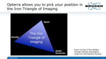
Opterra allows you to pick your position in the Iron Triangle of Imaging Figure courtesy of Paul Maddox Concept originally developed by Jonas Dorn and Gaudenz Danuser 10/30/2014
Open the catalog to page 3
Opterra Design Overview • Two modes of operation • Variable pinhole, linear array field scanner. Supravideo rate, high speed slit scanner. • EMCCD camera-based detector. • High frequency scanning minimizes phototoxicity and photobleaching • Multiple scan modes allow for optimization of resolution, speed and light delivery • Linear pinholes and separate light paths for emission and excitation minimize emission crosstalk
Open the catalog to page 4
Overhead View of Opterra Imaging Scanner Showing Lightpath Bruker Confidential
Open the catalog to page 5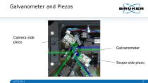
Camera side piezo Galvanometer Scope side piezo
Open the catalog to page 6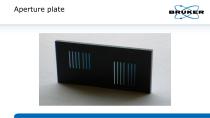
Aperture plate
Open the catalog to page 7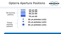
Opterra Aperture Positions Slit scanning positions 22 mm slit 35 mm slit 50 mm slit 70 mm slit 30 mm pinholes (x32) Pinhole scanning positions
Open the catalog to page 8
Opterra Imaging Scanner Light Path_ PINHOLE ARRAY From laser
Open the catalog to page 9
Time to move depends on exposure time It takes 2 frames for one complete “piezo sweep” Galvo sweeps multiple cycles/frame, and piezos sweep only once per frame Time to move depends on exposure time Piezos move a fixed distance Galvo moves a fixed distance Galvanometer and Piezo Movement
Open the catalog to page 10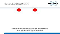
Galvanometer and Piezo Movement Field-scanning combines multiple galvo sweeps with bidirectional piezo movement
Open the catalog to page 11
Scan pattern timing Sample Piezo/Galvo/Blanking Waveforms For Single Frame piezo offset galvo offset piezo waveform galvo waveform AOTF blanking (R)est, (A)ccelerate, (F)ront porch, (S)canning, (B)ack porch, (D)ecelerate
Open the catalog to page 12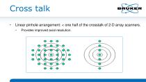
Cross talk • Linear pinhole arrangement: < one half of the crosstalk of 2-D array scanners. • Provides improved axial resolution.
Open the catalog to page 13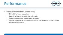
Standard Opterra camera (Evolve Delta) • 512 x 512 full frame acquisition. Up to 40 full frames per second pinhole mode. Faster acquisition from smaller region of interest. Slit scan imaging at 66 fps full frame (512x512), 500 fps with ROI, up to 1000 fps with specialized detector
Open the catalog to page 14
Opterra benefits for Multidimensional Imaging • Less cross talk provides improved axial resolution and imaging depth • High acquisition speed provides excellent temporal resolution • Shorter exposure times results in lower phototoxicity and photobleaching • Sea urchin embryos, 50 plane Z series every 2 minutes, experiments for over 2 hours compare to 1 hour on spinning disc Generally across a number of samples exposures 5 – 10 times shorter than spinning discs
Open the catalog to page 15
Zebrafish Neurons 100 uM Z Range Opterra confocal
Open the catalog to page 16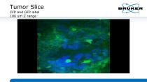
Tumor Slice CFP and GFP label 100 um Z range
Open the catalog to page 17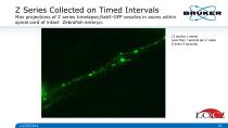
Z Series Collected on Timed Intervals Max projections of Z series timelapse,Rab5-GFP vesicles in axons within spinal cord of intact Zebrafish embryo. -. 12 section z series Less than 1 second per Z stack Z every 5 seconds
Open the catalog to page 18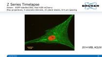
Green: EGFP labelled EB3, Red H2B mCherry Max projections, 5 seconds intervals, 21 plane stacks, 0.5 um spacing
Open the catalog to page 19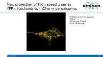
Max projection of high speed z series YFP mitochondria, mCherry peroxisomes Max projection of high speed z series 11 Planes, 0.3 micron spacing 2 Color 1.2 seconds / Z stack Z every 2 seconds
Open the catalog to page 20
Atlas Imaging for Stage Montages
Open the catalog to page 21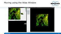
Moving using the Atlas Window BRUKER
Open the catalog to page 22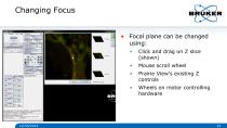
Changing Focus • Focal plane can be changed using: • • Mouse scroll wheel Prairie View’s existing Z controls Click and drag on Z slice (shown) Wheels on motor controlling hardware
Open the catalog to page 23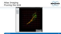
Atlas Imaging BRUKEI Pruning the Grid (JXJ>
Open the catalog to page 24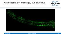
Arabidopsis 2x4 montage, 60x objective BRUKER Bruker Confidential
Open the catalog to page 25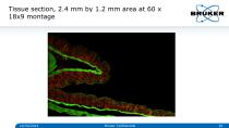
Tissue section, 2.4 mm by 1.2 mm area at 60 x 18x9 montage Bruker Confidential
Open the catalog to page 26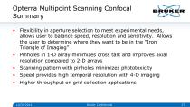
Opterra Multipoint Scanning Confocal Summary • Flexibility in aperture selection to meet experimental needs, allows user to balance speed, resolution and sensitivity. Allows the user to determine where they want to be in the “Iron Triangle of Imaging” • Pinholes in 1-D array minimizes cross talk and improves axial resolution compared to 2-D arrays • Scanning pattern with pinholes minimizes phototoxicity • Speed provides high temporal resolution with 4-D imaging • Higher throughput on grid collection applications Bruker Confidential
Open the catalog to page 27
© Copyright Bruker Corporation. All rights reserved.
Open the catalog to page 28Archived catalogs
 Innova
8 Pages
 Dimension FastScan
8 Pages
 MultiMode 8 Brochure
8 Pages
 Dimension Icon
6 Pages
 Dimension FastScan Bio
4 Pages
 AFM Dimension Edge
8 Pages
 BioScope Resolve
8 Pages
 microflex
6 Pages
 MALDI PharmaPulse HTS
4 Pages
 The new autoflex speed
10 Pages
 rapifleX MALDI Tissuetyper
8 Pages
 PRIME
PRIME12 Pages
 MALDI Biotyper CA System
8 Pages
 MBT STAR-BL Software
4 Pages
 AutoMet AFM Software
2 Pages
 Dimension Icon SSRM
2 Pages
 MultiMode 8-HR
8 Pages


- BRUKER microscope
- BRUKER optical microscope
- BRUKER laboratory microscope
- BRUKER benchtop microscope
- Spectroscope
- BRUKER biology microscope
- Benchtop spectrometer
- Fluorescence microscope
- BRUKER research microscope
- Digital microscope
- BRUKER high-resolution microscope
- Medical microscope
- Compact microscope
- Inverted microscope
- Zoom microscope
- Optical spectrometer
- Research spectrometer
- 3D microscope
- Confocal microscope
- High-resolution spectrometer