- Catalogs
- Bruker Nano Surfaces
- Opterra Multipoint Scanning Confocal Microscope
- Products
- Catalogs
- News & Trends
- Exhibitions
Opterra Multipoint Scanning Confocal Microscope

Opterra Multipoint Scanning Confocal Microscope
Overview
The Bruker Opterra Multipoint Scanning Confocal Microscope is engineered for live-cell fluorescence microscopy, combining high resolution typical of confocal systems with the speed of wide-field imaging. It uses a CCD camera detector and offers selectable aperture sizes to optimize speed and resolution for diverse experimental needs. Its unique optical design enables spectral imaging of dynamic processes with minimal photobleaching and phototoxicity, making it ideal for advanced live-cell studies such as protein localization, intracellular ion imaging, and cellular dynamics.
Key Features and Performance
- High-speed, multidimensional imaging of live cells and small organisms.
- Photoactivation module tailored for cellular kinetic studies.
- Intuitive, powerful Prairie View software for complex imaging protocols.
- Motorized aperture plate with 7 selectable apertures (3 pinholes and 4 slits) allowing optimization between resolution and speed without hardware changes.
- One-dimensional pinhole arrays reduce crosstalk compared to two-dimensional spinning disk arrays, resulting in sharper images with better resolution and imaging depth.
- Spectral imaging at 4 frames per second with 15 spectral channels per frame at 512x512 pixel resolution.
- Simultaneous photoactivation and imaging without emission signal loss.
Comparison with Spinning Disk Systems
The Opterra system offers advantages over traditional spinning disk confocals, including:
- Higher speed operation with slit apertures (up to 1000 fps).
- Reduced photobleaching and phototoxicity.
- Improved axial resolution and imaging depth due to linear pinhole configuration.
- Software-controlled aperture selection without hardware modifications.
- Enhanced spectral imaging capabilities.
- Integrated photoactivation with no emission loss, supporting advanced photomanipulation techniques.
Software and Data Acquisition
Powered by Prairie View software, the system supports:
- User-friendly setup of time lapse, Z-series, stage montages, and multidimensional acquisitions.
- Integration of external devices via triggers and analog signals.
- Atlas Imaging module for simplified multifield and stage montage imaging with intuitive navigation in X, Y, and Z.
- Real-time spectral data processing and visualization.
- Control of photostimulation protocols including points, lines, rectangles, arbitrary shapes, and spiral activation.
- Support for complex photoactivation experiments such as FRAP, photoconversion, optogenetics, and uncaging.
Applications
Typical applications include:
- Protein localization and trafficking
- Vesicle trafficking
- Mitosis and microtubule dynamics
- Organelle trafficking
- Cell migration
- Intracellular ion concentration measurements
- FRAP and photoconversion
- Cell membrane wound healing
- Photoactivation and optogenetics
Optical and Mechanical Specifications
- Scanning method: Combination of galvanometer and piezoelectric crystal scanning.
- Apertures: Motorized plate with 3 pinhole sizes (30, 45, 60 µm) and 4 slit widths (22, 35, 50, 70 µm).
- Scan speeds: Up to 50 fps in pinhole mode; up to 1000 fps in slit mode with appropriate camera.
- Filters: Motorized 6-position emission filter wheel; custom dichroics and polychroics.
- Triggering: Frame trigger in/out and 4 auxiliary device triggers.
- Detector: CCD camera with support for Photometrics and Q-Imaging cameras; standard Evolve Delta EMCCD or Rolera EM-C2 EMCCD.
- Optical inputs: Helios launch with up to 5 diode laser lines, analog modulated with digital blanking; fiber optic input.
- Microscope platforms: Nikon TI-E inverted and Nikon FN-1 upright with software control of turret and Z-motor.
- Specimen stage: ASI XY stage with piezo focus insert (300 µm range) for TI-E; Bruker Slim Stage for FN-1.
- Z focus: Internal focus motor and piezo for TI-E; Bruker Z-focus motor and piezo-driven objective focus for NI-E.
- Photoactivation scanner: Single-pair galvanometer (3 mm) for high-speed photoactivation, photobleaching, and ablation.
- Analog I/O: Up to 8 analog outputs and 8 analog inputs for device control and signal recording.
- Software: Prairie View Imaging Software on Windows 7 64-bit.
- Computer: Intel Quad Core processor, multiple SSDs for OS, software, and data, 32GB RAM.
Summary
The Bruker Opterra Multipoint Scanning Confocal Microscope is a versatile, high-performance imaging platform optimized for live-cell fluorescence microscopy. Its unique optical design, flexible aperture selection, and integrated photoactivation capabilities provide superior imaging speed, resolution, and spectral imaging compared to traditional spinning disk systems. Coupled with powerful and user-friendly software, it supports a wide range of advanced biological applications requiring dynamic, multidimensional imaging with minimal photodamage.
Catalog excerpts

Opterra Multipoint Scanning Confocal Microscope Cell-Friendly, High-Speed, High-Resolution Imaging Innovation with Integrity Fluorescence Microscopy
Open the catalog to page 1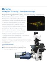
Multipoint Scanning Confocal Microscope Superior Integration, Versatility, and Cell-Friendly Performance The Opterra™ Multipoint Scanning Confocal Microscope is designed specifically for live-cell microscopy. It uniquely combines the resolution of traditional confocal systems with the speed typically associated with wide-field imaging. The system utilizes a CCD camera as a detector, and can be configured through selectable aperture sizes so the optimal balance of speed and resolution for each experiment can be set to meet your specific application requirements. Opterra’s unconventional optical...
Open the catalog to page 2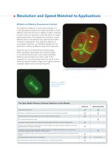
Resolution and Speed Matched to Applications Efficient and Effective Fluorescence Confocal One aspect of Opterra’s unique scanner design is a motorized aperture plate that contains pinholes of three different sizes and slits of four different widths. Pinholes provide maximum resolution, while slits allow for higher speed acquisitions. This enables the scientist to match aperture size to the objective and optimize the system for specific applications. Plus, aperture selection is controlled by software, so hardware changes are not required for setting up different experiment protocols. Opterra’s...
Open the catalog to page 3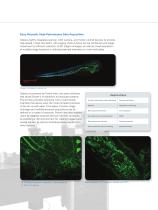
Easy, Versatile, High-Performance Data Acquisition Opterra tightly integrates scanner, CCD camera, and motion control devices to provide high-speed, timed volumetric, 4D imaging. Piezo Z-focus can be combined with stage movement for efficient collection of 3D stage montages, as well as timed acquisition at multiple stage locations in individual sample chambers or multi-well plates. Stage montage of zebrafish. Opterra is powered by Prairie View, the same software that drives Bruker’s multiphoton microscope systems. Prairie View provides scientists with a user-friendly interface that allows even...
Open the catalog to page 4
Flexible and Feature-Rich Software Multifield Imaging Simplified Prairie View’s Atlas Imaging module makes setting up stage montages and other types of multiple stage location paradigms simple and intuitive. Easy navigation in X, Y and Z allows the user to quickly find and record individual locations of interest, or define a grid of overlapping locations which can be used to construct a high-resolution image of up to an entire specimen. The preview window shows a coarse tiling of all positions scanned, and the live window shows the currently selected field of view. From the quick-scan preview,...
Open the catalog to page 5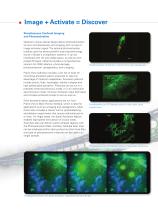
Image + Activate = Discover Simultaneous Confocal Imaging and Photoactivation Opterra’s unique optical design allows photostimulation to occur simultaneously with imaging with no loss of image emission signal. The optional photoactivation module uses the same powerful scanning technology found in Bruker’s multiphoton systems. It can be interfaced with UV and visible lasers, as well as with pulsed IR lasers. Opterra provides a comprehensive solution for FRAP ablation, photo-damage, , photoconversion, optogenetics, and uncaging. Photoconversion of Dendra in glioma cell using masked area. Prairie...
Open the catalog to page 6
Dynamic Spectral Scanning for Live Cell Imaging Opterra’s Spectral Scanning feature performs spectral imaging at speeds appropriate for live cell and small organism imaging. By incorporating an Amici prism into the Opterra image scanner, full spectral data sets of 15 channels can be acquired at speeds of up to four spectral data sets per second for a 512x512 pixel image. Prairie View software provides real time processing of spectral data sets to provide a live image in which up to four channels of data can be visualized. Spectral Scanning also can be used with time lapse, Z-series, and stage...
Open the catalog to page 7
Scanhead Scanning Method Combination galvanometer and piezoelectric crystal scanning Fully motorized aperture plate with 7 software-selectable apertures; 3 pinhole settings (30, 45, and 60 µm); 4 slit settings (22, 35, 50, and 70 µm) Scan Speeds Scanner or camera master timed imaging; Up to 50 fps in pinhole mode; Up to 1000 fps in slit imaging mode (with appropriate camera) Motorized 6-postion emission filter wheel; Custom dichroics and polychroics for multiple excitation/emission wavelengths Frame trigger out and frame trigger in; 4 auxillary device triggers Image Collection Camera port with...
Open the catalog to page 8Archived catalogs
 Innova
8 Pages
 Dimension FastScan
8 Pages
 MultiMode 8 Brochure
8 Pages
 Dimension Icon
6 Pages
 Dimension FastScan Bio
4 Pages
 AFM Dimension Edge
8 Pages
 BioScope Resolve
8 Pages
 microflex
6 Pages
 MALDI PharmaPulse HTS
4 Pages
 The new autoflex speed
10 Pages
 rapifleX MALDI Tissuetyper
8 Pages
 PRIME
PRIME12 Pages
 MALDI Biotyper CA System
8 Pages
 MBT STAR-BL Software
4 Pages
 AutoMet AFM Software
2 Pages
 Dimension Icon SSRM
2 Pages
 MultiMode 8-HR
8 Pages


- Radiology Support Devices optical microscope
- Radiology Support Devices laboratory microscope
- Radiology Support Devices benchtop microscope
- Spectroscope
- Radiology Support Devices biology microscope
- Benchtop spectrometer
- Fluorescence microscope
- Radiology Support Devices research microscope
- Digital microscope
- Radiology Support Devices high-resolution microscope
- Medical microscope
- Compact microscope
- Inverted microscope
- Zoom microscope
- Optical spectrometer
- Research spectrometer
- 3D microscope
- High-resolution spectrometer