- Catalogs
- Caliber Imaging & Diagnostics
- VIVASCOPE® 3000
VIVASCOPE® 3000
1 /4Pages

VIVASCOPE® 3000
1 /4Pages
Catalog excerpts

non-invasive cellular imaging of the skin
Open the catalog to page 1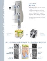
CONFOCAL IMAGING VIVASCOPE® systems are in vivo confocal imaging tools that use a low-powered laser to provide non-invasive, real-time, highresolution images of the epidermis and the superficial collagen layers.* The system captures the natural reflectivity of different cellular structures that can offer important information that may be useful in forming a clinical judgment. Cross section of skin Confocal image stack HOW A CONFOCAL IMAGE CORRELATES TO A TRADITIONAL PERSPECTIVE RETICULAR DERMIS
Open the catalog to page 2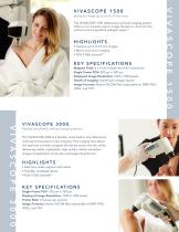
ability to image up to an 8 x 8 mm area The VIVASCOPE 1500 reflectance confocal imaging system offers a non-invasive way to image the skin in vivo from the surface to the superficial collagen layers.* • Capture up to 8 x 8 mm images • Macro and micro imaging • FDA 510(k) cleared** Mapped Field: 8 x 8 mm in both the X & Y directions Single Frame FOV: 500 µm x 500 µm Displayed Image Resolution: 1024 x 1024 pixels Depth of Imaging: Superficial collagen layers* Image Formats: Native DICOM files exportable as: BMP, PNG, JPEG, and TIFF flexible hand-held confocal imaging device The VIVASCOPE 3000 is...
Open the catalog to page 3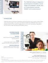
The VIVASCAN software is designed to provide physicians the ability to review images at the bedside or transfer them to a remote physician using the VIVANET communications system. V I VA S C A N VIVASCAN software incorporates comprehensive features that allow users to acquire, transfer, display, review, and store images, in one, easy-to-use dashboard. Additionally, VIVASCAN makes it easy to schedule patients for exams, perform imaging exams on one or more areas during a single patient visit, and retrieve images associated with the patient’s history. Areas of interest are identified by the patient...
Open the catalog to page 4All Caliber Imaging & Diagnostics catalogs and technical brochures
 CALIBER ID RS-G4
4 Pages
 VIVASCOPE 2500
2 Pages
 VivaScope
4 Pages
- Analysis software
- Microscopy
- Laboratory microscope
- Desktop microscope
- Visualization software
- Control software
- Laboratory software
- Windows software
- Cloud-based software
- Acquisition software
- Capture software
- Research microscope
- Sharing software
- Cell imaging system
- Interpretation software
- Automatic cell imaging system
- Zoom microscope
- Laboratory cell imaging system
- Confocal microscope
- Laser microscope