
Catalog excerpts
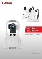
CR-2 AF/ CR-2 Plus AF Digital Retinal Camera Non-Mydriatic cameras
Open the catalog to page 1
Extremely compact and lightweight (just 15 kg) camera: suitable for mobile use. Non-Mydriatic camera with additional fundus autofluorescence (FAF) capability. Short reaching distance The compact design allows the operator to easily keep the patient’s eye open with one hand and permits an excellent view of the patient’s eye. Specially shaped surface to act as grip; easy handling for quick and efficient image capture. Multifunctional joystick Up and down movement of optical head (powered) and focus ring. For optimized viewing angles, so camera can be operated while seated or standing up.
Open the catalog to page 2
Extensive Auto Functions Auto Focus Fast and accurate automatic focusing. Auto Shot Once the alignment, working distance and focus are correct, photography is done automatically. Auto Switching from Anterior to Retina When aligned correctly on the pupil, the camera will automatically switch to retinal observation view. Photometric Auto Exposure Flash and observation light intensity is set automatically for every examination, based on retina reflectance, for perfect images regardless of pupil size or ethnicity. Full Control The auto functions will make the procedure much easier. Nevertheless...
Open the catalog to page 3
Photography Modes Color High quality 45 degrees image. 2 X digital magnification. Digital Redfree Digital Cobalt Digital Red Free and Digital Cobalt Automatically created from the original RAW color image. Based on the EOS retina technology and Canon proprietary image processing. Image quality comparable with optical filters. Anterior Photography Quick and easy anterior segment photography to document the cornea, pupil, eyelid and sclera.
Open the catalog to page 4
Fundus Autofluorescence (CR-2 Plus AF only) Macular blood FAF imaging for the diagnosis of retinal disease is a relatively new diagnostic technique that provides more information on the health of the retinal pigment epithelium. FAF has proven to be very useful for the early detection of Age-related Macular Degeneration (AMD), one of the leading causes of visual impairment. Recent studies indicate that FAF imaging can also aid the diagnosis of a variety of other diseases and even detection of intraocular tumors. AMD With the extra feature of FAF photography we have discovered retinal changes...
Open the catalog to page 5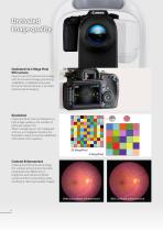
Unrivaled Image quality Dedicated 32.5 Mega Pixel EOS camera Canon’s own EOS camera technology, with its renowned image processing capabilities, is adapted exclusively for Canon retinal cameras, it provides optimal retinal imaging. Resolution Important factor that contributes to a high image quality is the number of pixels per square mm. When comparing our 32.5 megapixel with e.g. a 4 megapixel camera, the illustration clearly shows the additional information that is gained. Contrast Enhancement Utilizing the EOS retina technology, this contrast enhancement function emphasizes the...
Open the catalog to page 6
Canon Opacity Suppression When obtaining retinal images, ocular opacities will cause several problems: the scattering of the light will make the edges of the blood vessels appear blurred. The difference in brightness of the retina will be reduced, making it very difficult to distinguish between structures. And a cataract eye will cause images to appear more yellow. With Canon’s unique and sophisticated opacity suppression tool the blood vessels will appear much clearer; the original brightness of the retina will be restored and any change in retinal color will be compensated. With Canon...
Open the catalog to page 7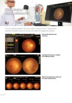
Canon Retinal Expert software RX The new software platform for Canon retinal cameras and OCT. Designed for seamless integration and connectivity with patient management systems. Extremely intuitive user interface Compare both eyes or studies from different dates Observe progression; select up to 5 past examinations
Open the catalog to page 8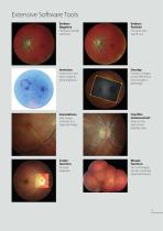
Extensive Software Tools Emboss Negative Emboss Positive The blood vessels stand out. The optic disc stands out. Inverts the color of an image to assist diagnosis. Overlay 2 images to see differences and changes in pathology. Cup/disc measurement Add shapes and texts to a captured image. Measure the optic nerve papillary area. Loupe function Mosaic function Up to 20 images can be combined (optional feature).
Open the catalog to page 9
Canon Retinal Expert Software Platform RX Stand alone configuration All-in one system. Capturing, viewing and database. RX Viewer • Reviewing • Reporting Optional RX Viewers can be connected over the network and access the database of the device. Up to 2 RX viewers can access the database at the same time. • Capturing • Reviewing and reporting • Database and archive • Reviewing • Reporting A Canon OCT could be added to the stand alone configuration, sharing the same PC and database. Network configuration With RX Server up to 5 systems can be connected with maximum 10 concurrent viewers. RX...
Open the catalog to page 10
Seamless Integration with Patient Management Systems The Canon RX software can automatically start the patient management software on the selected patient and vice versa. (Command Line Interface) Third party software can start the Canon RX software Practice management softPractice ware management RX Software shows data of that patient Command with patient information Canon RX software can start third party software RX Software Third party software opens on that patient 2 3 Program launch preset keys Command with patient information Versatile Patient data input possibilities for optimal...
Open the catalog to page 11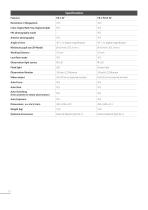
Color. Digital Red Free, Digital Cobalt Anterior photography Minimum pupil size (SP Mode) Working Distance Observation light source Flash light Strobo tube Observation Monitor Video output Full HD on an external monitor Full HD on an external monitor Auto Focus Auto Shot Auto Switching (from anterior to retina observation) Auto Exposure Optional Accessories External fixation light (EL-1) External fixation light (
Open the catalog to page 12
コンポジットロゴ_CANON MEDICAL SYSTEMS USA,INC_英語表記 To schedule a demo or for additional information, call (833) 521-3937 or visit our website. Canon Medical Systems USA, Inc. https://us.medical.canon | 2441 Michelle Drive, Tustin CA 92780 | 800.421.1968 ©Canon Medical Systems, USA 2023. All rights reserved. Design and specifications are subject to change without notice. Made for Life is a trademark of Canon Medical Systems Corporation. YouTube logo is a trademark of Google Inc. TWITTER, TWEET, RETWEET and the Twitter logo are trademarks of Twitter, Inc. or its affiliates. LinkedIn, the LinkedIn...
Open the catalog to page 13All Canon Medical System U.S.A catalogs and technical brochures
-
CX-1
13 Pages
-
Aquilion ONE / PRISM Edition
26 Pages
-
Aplio a550
16 Pages
Archived catalogs
-
RK-F2
4 Pages
-
CR-2 PLUS AF
8 Pages
-
OMNERA® 400A
2 Pages
-
CXDI-50RF Specifications
1 Pages
-
CR-2 AF
8 Pages
-
Aplio a550
20 Pages
-
Aplio a450
16 Pages
-
Vantage Titan
17 Pages
-
Vantage Elan
19 Pages
-
Vantage Galan 3T
40 Pages
-
RadPRO® URS
2 Pages
-
RadPRO® OMNERA® 400T
2 Pages
-
CXDI-801C
1 Pages
-
CXDI-701C
1 Pages
-
CXDI-401C
1 Pages
-
flyer RICS
2 Pages
-
image SPECTRUM Brochure
8 Pages
-
CR2 Plus AF Brochure
8 Pages
-
RK-F2
2 Pages
-
PTS 1000
2 Pages
-
TX-20
2 Pages
-
CR-2 PLUS
2 Pages
-
CR-2
2 Pages
-
CF-1
2 Pages
-
CX-1
4 Pages
-
CXDI-60G
4 Pages
-
CXDI-60C
2 Pages
-
CXDI-55G
2 Pages
-
CXDI-55C
2 Pages
-
CXDI-401 COMPACT
2 Pages
-
CXDI-401
2 Pages
-
CXDI-501G
2 Pages
-
CXDI-501C
2 Pages
-
CXDI-80C
2 Pages
-
CXDI-70C Wireless
8 Pages


