- Catalogs
- Canon Medical System U.S.A
- CR-2 PLUS AF
CR-2 PLUS AF
1 /8Pages

CR-2 PLUS AF
1 /8Pages
Catalog excerpts

CR-2 PLUS AF DIGITAL NON-MYDRIATIC RETINAL CAMER A AutoFunctionality Plus Fundus AutoFluorescence
Open the catalog to page 1
CR-2 PLUS AF SEE PRECISELY WHAT YOU’VE BEEN MISSING INTEGRATED TECHNOLOGY The Canon CR-2 PLUS AF Digital Retinal Camera is designed to help you consistently capture and analyze truly superb images—quickly, efficiently, and automatically. Designed around the legendary Canon EOS optics and advanced CMOS image capture technology, the CR-2 PLUS AF Digital Retinal Camera provides a remarkable set of advanced features specifically designed to enhance, capture, and analyze all fundus images. DEDICATED DIGITAL CAMERA Canon is the only brand offering a digital fundus camera that incorporates its own premium...
Open the catalog to page 2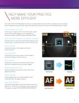
HELP MAKE YOUR PRACTICE MORE EFFICIENT The Canon CR-2 PLUS AF Digital Retinal Camera is extremely flexible and comes with a comprehensive set of standard features designed to help you quickly capture and analyze data. Some of the camera’s more noteable features include: AUTOFLUORESCENCE (FAF) At the touch of a button, the CR-2 PLUS AF provides instant access to this valuable diagnostic tool, enabling you to document changes in the Retinal Pigment Epithelium (RPE). AUTO-FOCUS WITH MANUAL ALIGNMENT OVERRIDE Automatically focus the eye by partially depressing the joystick, or easily switch to manual...
Open the catalog to page 3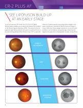
CR-2 PLUS AF SEE LIPOFUSCIN BUILD-UP AT AN EARLY STAGE In the non-mydriatic FAF mode, the CR-2 PLUS AF Digital Retinal Camera enables you to assess and monitor the condition of the Retinal Pigment Epithelium (RPE). The camera’s infrared alignment light allows patients with pupils as small as 3.3 mm to be easily photographed without dilation drops. FAF is especially effective in identifying and monitoring subtle changes in the areas of lipofusion, even before there’s structural change or vision is affected. These changes may be pre-cursors to vision problems, such as diabetic retinopathy, age-related...
Open the catalog to page 4
COUNT ON EXTENSIVE DIGITAL FILTER PROCESSING CAPABILITIES The CR-2 PLUS AF Digital Retinal Camera produces ultrahigh-resolution, wide-angle views with excellent color, detail, and contrast. To further enhance your retinal exam capabilities, the camera has a full set of blue, green, and red digital processing modes to extract more in-depth information from each image. CHANNEL MODES COLOR Excellent views of the retinal nerve fiber layer, internal limiting membrane, retina folds, cysts, and epiretinal membranes. Useful for pigmentary disturbances, choroidal ruptures, choroidal nevi, and choroidal...
Open the catalog to page 5
CR-2 PLUS AF imageSPECTRUM IMAGE MANAGEMENT SOFTWARE ADVANCED IMAGE ANALYSIS DATA PORTABILITY EFFICIENT IMAGE MANAGEMENT UNIVERSAL IMAGE CAPTURE DICOM-BASED STANDARD PATIENT HEALTH INFORMATION SECURITY FEATURES ADVANCED IMAGE ANALYSIS Use Advanced Tools to help quantify progression; overlay and merge images to clearly see and assess changes; add notes to help document and annotate. DATA PORTABILITY Provides for the secure transmission of patient data to referring physicians and other coordinated care partners. EFFICIENT IMAGE MANAGEMENT This is not an EMR. It’s software specifically designed...
Open the catalog to page 6
imageSPECTRUM FITS EASILY INTO NEARLY ANY BUSINESS ENVIRONMENT imageSPECTRUM can be easily configured to support virtually any practice network, small or large. Designed with your growing practice in mind, this system is readily scalable. This means that imageSPECTRUM can support not only the needs of your current practice, but also easily expand as your practice grows or your needs change. A SINGLE-SITE NETWORK WITH MULTIPLE EXAM ROOMS Instrument Stations Data Storage Reception Area Exam Rooms Physician’s Office Office #3 Office #4 A MULTISITE NETWORK WITH MULTIPLE EXAM ROOMS
Open the catalog to page 7
DIGITAL NON-MYDRIATIC RETINA CAMERA Specifications Canthus Mark: 420 mm From Base Type: Digital Retinal Camera, Non-Mydriatic Internal Eye Fixation: LED Dot Matrix Type of Photography Color, Digital Red-free, Digital Cobalt, Fundus AutoFluorescence (FAF) External Eye Fixation: White LED (Sold Separately) Angle of View: 45º (35º SP Mode) Working Distance Adjustment Anterior Observation: Double Image Match Method Fundus Observation: Working Distance Dots Minimal Pupil Size: 4.0 mm (3.3 mm SP mode) Sensor Resolution: 18.0 megapixels or more Focus Adjustment Type: Split-Line Adjustment Camera Dedicated...
Open the catalog to page 8All Canon Medical System U.S.A catalogs and technical brochures
 CX-1
CX-113 Pages
 Aquilion ONE / PRISM Edition
26 Pages
 CR-2 AF/ CR-2 Plus AF
13 Pages

Archived catalogs
 CXDI-70C Wireless
8 Pages
 CXDI-80C
2 Pages
 CXDI-501C
2 Pages
 CXDI-501G
2 Pages
 CXDI-401
2 Pages
 CXDI-401 COMPACT
2 Pages
 CXDI-55C
2 Pages
 CXDI-55G
2 Pages
 CXDI-60C
2 Pages
 CF-1
CF-12 Pages
 CXDI-60G
4 Pages
 CR-2
CR-22 Pages
 CR-2 PLUS
2 Pages
 TX-20
TX-202 Pages
 RK-F2
RK-F22 Pages
 PTS 1000
2 Pages
 CR2 Plus AF Brochure
8 Pages
 image SPECTRUM Brochure
8 Pages
 flyer RICS
2 Pages
 CXDI-401C
1 Page
 CXDI-701C
1 Page
 CXDI-801C
1 Page
 RadPRO® OMNERA® 400T
2 Pages
 RadPRO® URS
2 Pages
 Vantage Galan 3T
40 Pages
 Vantage Elan
19 Pages
 Vantage Titan
17 Pages
 Aplio a450
16 Pages
 Aplio a550
20 Pages
 CR-2 AF
8 Pages
 CXDI-50RF Specifications
1 Page
 OMNERA® 400A
2 Pages
 RK-F2
RK-F24 Pages

- Analysis software
- Ultrasound system
- B/W ultrasound system
- Color doppler ultrasound system
- Multipurpose ultrasound imaging system
- Visualization software
- Radiology software
- Tablet computer software
- Tablet PC software
- Flat panel detector
- Control software
- Digital radiography system
- Windows software
- Convex-array ultrasound system
- Linear-array ultrasound system
- Multipurpose radiography system
- X-ray system
- Multipurpose radiography X-ray system
- On-platform ultrasound system
- Portable flat panel detector