CX-1
1 /13Pages

CX-1
1 /13Pages
Catalog excerpts
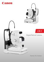
CX-1 Digital Retinal Camera
Open the catalog to page 1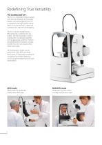
Redefining True Versatility The multifaceted CX-1 The CX-1 is a Mydriatic Retinal Camera with full Non-Mydriatic functionality. Besides color photography, the CX-1 is equipped with high quality optical filters for FLUO, Red Free, Cobalt and standard even with FAF photography. The CX-1 can be changed into a NM camera by a simple push of a button. The Non-Mydriatic mode is essential for non dilatable patients such as glaucoma suspects. Children and photosensitive patients will also benefit from the non invasive IRED observation light. All photography modes can be performed in the MYD and NON MYD...
Open the catalog to page 2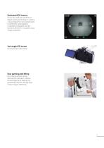
Dedicated EOS camera Canon has used their expertise in digital camera technology to create a unique digital EOS camera dedicated to ophthalmic photography: Completely integrated with the functions of the CX-1 to assist in easy image acquisition. Vari-angle LCD screen For ergonomic observation. Easy panning and tilting For working around central obstructions (cataracts, vitreous hemorrhages) and imaging the peripheral retina for creating large mosaic images effortlessly.
Open the catalog to page 3
Easy Operation Compact device For maximal patient interaction. Easy to observe patient. Short reaching distance for easy opening patient’s eyelid. Motorized filters; easy to operate and protected from dust. Motorized chin rest for easy adjustment. Automatically optimized flash range The CX-1 has an automatically optimized flash range, adjusted to the different photography modes and ISO settings.
Open the catalog to page 4
Extensive Photography Modes Sophisticated Optical Filters for Highest Image Quality All photography modes are available in MYD or NON-MYD mode. Base Line Visualizing Nerve Fiber Layer, important for checking for Glaucoma Checking retinal flow for occlusions and leakages Useful for checking the condition of the blood vessels, important for detecting Diabetic Retinopathy FAF FAF Imaging for the diagnosis of retinal disease is a relatively new diagnostic technique that provides more information on the health of the retinal pigment epithelium. FAF has proven to be very useful for the early detection...
Open the catalog to page 5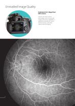
Unrivalled Image Quality Dedicated 32.5 Mega Pixel EOS camera Canon’s own EOS camera technology, with its renowned image processing capabilities, is adapted exclusively for Canon retinal cameras, it provides optimal retinal imaging.
Open the catalog to page 6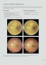
Canon Opacity Suppression When obtaining retinal images, ocular opacities can cause several problems. Canon opacity suppression tool is a unique and sophisticated software tool, that based on information from the EOS CMOS sensor will largely suppress the effect of ocular opacities on color images. Ocular opacities With Canon Opacity Suppression • The scattering of the light will make the edges of the blood vessels appear blurred • The difference in brightness of the retina will be reduced, making it very difficult to distinguish between structures • A cataract eye will cause images to appear...
Open the catalog to page 7
Canon Retinal Expert software RX The new software platform for Canon retinal cameras and OCT. Designed for seamless integration and connectivity with patient management systems. Extremely intuitive user interface Compare both eyes or studies from different dates Observe progression; select up to 5 past examinations
Open the catalog to page 8
Extensive Software Tools Emboss Negative Emboss Positive The blood vessels stand out. The optic disc stands out. Inverts the color of an image to assist diagnosis. Overlay 2 images to see differences and changes in pathology. Cup/disc measurement Add shapes and texts to a captured image. Measure the optic nerve papillary area. Loupe function Mosaic function Up to 20 images can be combined (optional feature).
Open the catalog to page 9
Canon Retinal Expert Software Platform RX Stand alone configuration All-in one system. Capturing, viewing and database. RX Viewer • Reviewing • Reporting Optional RX Viewers can be connected over the network and access the database of the device. Up to 2 RX viewers can access the database at the same time. • Capturing • Reviewing and reporting • Database and archive • Reviewing • Reporting A CX-1 could be added to a Canon OCT in a standalone configuration, sharing the same PC and database. Analysis results of both devices can be combined in one combined report. Network configuration With RX Server...
Open the catalog to page 10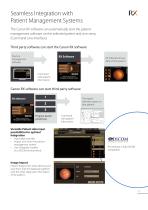
Seamless Integration with Patient Management Systems The Canon RX software can automatically start the patient management software on the selected patient and vice versa. (Command Line Interface) Third party software can start the Canon RX software Practice management softPractice ware management RX Software shows data of that patient Command with patient information Canon RX software can start third party software RX Software Third party software opens on that patient 2 3 Program launch preset keys Command with patient information Versatile Patient data input possibilities for optimal integration...
Open the catalog to page 11
Focus Adjustment Split Lines MYD: 50 degrees, Non-Myd: 45 degrees 2 X magnification (digital) Working distance Minimum pupil size Myd: ø 5.1mm (SP mode ø 4.3 mm) Panning and Non-Myd: ø 4.3 mm (SP mode ø 3.8 mm) Tilting Corneal Reflection dots adjustment 30 degrees to the left and right tilting range 15 degrees up, 10 degrees down Light sources Xenon tube for photography Halogenlamp for observation (Myd mode) IRED LED for observation (Non-Myd mode) Photography modes Mounted camera Dedicated digital EOS camera (32.5 MegaPixel for current model) HDMI Output for external monitor Full HD resolution...
Open the catalog to page 12
コンポジットロゴ_CANON MEDICAL SYSTEMS USA,INC_英語表記 To schedule a demo or for additional information, call (833) 521-3937 or visit our website. Canon Medical Systems USA, Inc. https://us.medical.canon | 2441 Michelle Drive, Tustin CA 92780 | 800.421.1968 ©Canon Medical Systems, USA 2023. All rights reserved. Design and specifications are subject to change without notice. Made for Life is a trademark of Canon Medical Systems Corporation. YouTube logo is a trademark of Google Inc. TWITTER, TWEET, RETWEET and the Twitter logo are trademarks of Twitter, Inc. or its affiliates. LinkedIn, the LinkedIn logo,...
Open the catalog to page 13All Canon Medical System U.S.A catalogs and technical brochures
 Aquilion ONE / PRISM Edition
26 Pages
 CR-2 AF/ CR-2 Plus AF
13 Pages

Archived catalogs
 CXDI-70C Wireless
8 Pages
 CXDI-80C
2 Pages
 CXDI-501C
2 Pages
 CXDI-501G
2 Pages
 CXDI-401
2 Pages
 CXDI-401 COMPACT
2 Pages
 CXDI-55C
2 Pages
 CXDI-55G
2 Pages
 CXDI-60C
2 Pages
 CF-1
CF-12 Pages
 CXDI-60G
4 Pages
 CR-2
CR-22 Pages
 CR-2 PLUS
2 Pages
 TX-20
TX-202 Pages
 RK-F2
RK-F22 Pages
 PTS 1000
2 Pages
 CR2 Plus AF Brochure
8 Pages
 image SPECTRUM Brochure
8 Pages
 flyer RICS
2 Pages
 CXDI-401C
1 Page
 CXDI-701C
1 Page
 CXDI-801C
1 Page
 RadPRO® OMNERA® 400T
2 Pages
 RadPRO® URS
2 Pages
 Vantage Galan 3T
40 Pages
 Vantage Elan
19 Pages
 Vantage Titan
17 Pages
 Aplio a450
16 Pages
 Aplio a550
20 Pages
 CR-2 AF
8 Pages
 CXDI-50RF Specifications
1 Page
 OMNERA® 400A
2 Pages
 CR-2 PLUS AF
8 Pages
 RK-F2
RK-F24 Pages

- Analysis software
- Ultrasound system
- B/W ultrasound system
- Color doppler ultrasound system
- Multipurpose ultrasound imaging system
- Visualization software
- Radiology software
- Tablet computer software
- Tablet PC software
- Flat panel detector
- Control software
- Digital radiography system
- Windows software
- Convex-array ultrasound system
- Linear-array ultrasound system
- Multipurpose radiography system
- X-ray system
- On-platform ultrasound system
- Multipurpose radiography X-ray system
- Portable flat panel detector