
- Catalogs
- Canon Medical Systems Europe
- https://eu.medical.canon/products/computed-tomography/piqe/


- Company
- Products
- Catalogs
- News & Trends
- Exhibitions
https://eu.medical.canon/products/computed-tomography/piqe/
https://eu.medical.canon/products/computed-tomography/piqe/
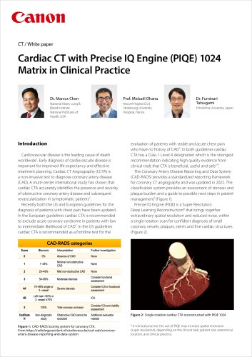
Cardiovascular disease is a leading cause of death globally, necessitating early diagnosis for effective treatment. Cardiac CT Angiography (CCTA) is recommended for diagnosing coronary artery disease (CAD) in patients with chest pain. The document discusses the use of PIQE 1024, a high-resolution imaging technique, in cardiac CT angiography, highlighting its clinical benefits and results from case studies.
PIQE is a Super Resolution Deep Learning Reconstruction technology that enhances spatial resolution and reduces noise in cardiac CT scans. It provides twice the resolution of conventional CT scans without additional radiation, aiding in the diagnosis of small coronary vessels, plaques, stents, and fine cardiac structures.
PIQE 1024 is beneficial for various cardiac applications, including Transaortic Valve Replacement (TAVR) planning. It offers higher spatial resolution and lower image noise, facilitating clearer visualization of small atherosclerotic plaques and calcifications. It improves CAD-RADs scoring and visualization of coronary arteries, stents, and prosthetic valves.
Different scan parameters are detailed, such as full beat volume scans with varying kV and CTDI vol., resulting in effective doses ranging from 2.48 mSv to 5.5 mSv.
Case studies demonstrate PIQE 1024's clinical benefits, including improved visualization of stenosis, plaque burden, and positive remodeling. Examples include:
- Improved lumen patency assessment in an 82-year-old with severe coronary calcifications.
- Clearer stent visualization in a 60-year-old man with an RCA stent.
- Sharper stent struts in a 69-year-old post-PCI.
- Better visualization of post-op calcifications in a 68-year-old with a mechanical aortic valve.
- Excellent delineation of aortic valve leaflets in a 64-year-old with an aortic aneurysm.
Studies support PIQE 1024 and SR-DLR's efficacy in improving image quality, reducing noise, and enhancing diagnostic accuracy in coronary CT angiography.
SR-DLR improves spatial resolution, reduces image noise, and enhances diagnostic confidence in evaluating coronary artery diseases. It is effective in identifying minimal non-obstructive coronary atherosclerosis and evaluating coronary stents.
To maximize SR-DLR's capabilities, it is recommended to reconstruct images with a 1024 matrix size, enhancing visualization of fine structures such as plaques in coronary arteries.
PIQE 1024 and SR-DLR significantly improve cardiac CT imaging, offering enhanced resolution and diagnostic value without additional radiation. They are valuable for assessing coronary artery disease and identifying vulnerable plaques, contributing to better patient outcomes.
Catalog excerpts
Cardiac CT with Precise IQ Engine (PIQE) 1024 Matrix in Clinical Practice Dr. Marcus Chen Prof. Mickaël Ohana National Heart, Lung & Blood Institute National Institutes of Health, USA Nouvel Hopital Civil, Strasbourg University Hospital, France Introduction Cardiovascular disease is the leading cause of death worldwide1. Early diagnosis of cardiovascular disease is important for improved life expectancy and effective treatment planning. Cardiac CT Angiography (CCTA) is a non-invasive test to diagnose coronary artery disease (CAD). A multi-center international study has shown that cardiac CTA accurately identifies the presence and severity of obstructive coronary artery disease and subsequent revascularization in symptomatic patients2. Recently both the US and European guidelines for the diagnosis of patients with chest pain have been updated. In the European guidelines cardiac CTA is recommended to exclude acute coronary syndrome in patients with low to intermediate likelihood of CAD3. In the US guidelines cardiac CTA is recommended as a frontline test for the Dr. Fuminari Tatsugami Hiroshima University, Japan evaluation of patients with stable and acute chest pain who have no history of CAD4. In both guidelines cardiac CTA has a Class 1 Level A designation which is the strongest recommendation indicating high quality evidence from clinical trials that CTA is beneficial, useful and safe3,4. The Coronary Artery Disease Reporting and Data System (CAD-RADS) provides a standardized reporting framework for coronary CT angiography and was updated in 2022. The classification system provides an assessment of stenosis and plaque burden and a guide to possible next steps in patient management5 (Figure 1). Precise IQ Engine (PIQE) is a Super Resolution Deep Learning Reconstruction* that brings together extraordinary spatial resolution and reduced noise, within a single-rotation scan for confident diagnosis of small coronary vessels, plaques, stents and fine cardiac structures (Figure 2). Figure 2: Single rotation cardiac CTA reconstructed with PIQE 1024 Figure 1: CAD-RADS Scoring system for coronary CTA. From https://radiologyassistant.nl/cardiovascular/cad-rads/coronaryartery-disease-reporting-and-data-system * In clinical practice, the use of PIQE may increase spatial resolution (super resolution), depending on the clinical task, patient size, anatomical location, and clinical practice.
Open the catalog to page 1
The PIQE Deep Learning Reconstruction algorithm is trained using Ultra-High Resolution data with twice the resolution of conventional CT acquired on the commercially available Aquilion Precision CT system, which features UHR 0.25 mm detectors in routine clinical practice. Datasets reconstructed with PIQE empower the clinician with twice the high contrast signal definition, as well as reduced noise, in all three dimensions, relative to conventional hybrid iterative reconstruction. These benefits maintain low contrast detectability, without additional radiation dose to the patient6 (Figure 3)....
Open the catalog to page 2
PIQE 1024 Matrix training principles The PIQE reconstruction algorithm features a next generation, three-dimensional neural network trained to identify and preserve signal features, both in-plane and longitudinally, throughout the cardiac volume dataset. Trained on high quality cardiac cases acquired on clinically operating Aquilion Precision systems, PIQE optimizes spatial resolution for clinically relevant tasks and realistic Training Data Preparation High Resolution CT Acquisition Model Aquilion Precision Anatomical Model Scanner Model field-of-views. PIQE’s three-dimensional learning also...
Open the catalog to page 3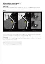
Stenosis in the right coronary artery (RCA) Dr. Tatsugami, Hiroshima University, Japan Patient History This 73-year-old man presented with typical angina pectoris and a heart rate of 60 bpm. A coronary CTA was requested. A mild stenosis of CAD-RADs 2 (25-49%) is seen in the proximal segment of the right coronary artery. Non-calcified plaque is identified at the stenotic site. A small calcification is seen immediately distal to the stenosis. Clinical Benefit The spotty calcification are described as high risk plaque features. PIQE 1024 may provide identification of micro calcifications beyond...
Open the catalog to page 4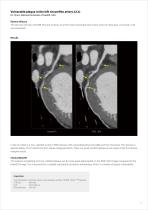
Vulnerable plaque in the left circumflex artery (LCx) Dr. Chen, National Institutes of Health, USA Patient History This 60-year-old man with BMI 29.4 and a history of an RCA stent presented with recent onset of chest pain. A Coronary CTA was requested. In the LCx there is a non-calcified severe (>70%) stenosis with outward/positive remodeling of the mid vessel. This stenosis is approximately 10 mm before the first obtuse marginal branch. There is a small, calcified plaque at the origin of the first obtuse marginal vessel. Clinical Benefit The positive remodeling and non-calcified plaque can be...
Open the catalog to page 5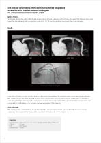
Left anterior descending artery (LAD) non-calcified plaque and correlation with invasive coronary angiogram Prof. Ohana, Strasbourg University Hospital, France Patient History This 67-year-old woman with a BMI 28 and a heart rate of 76 bpm presented with a history of angina. The Calcium Score scan was within normal range with an Agatston score of 68. A CTA was requested to investigate the cause of angina. Invasive coronary angiogram In the mid LAD there is a non-calcified plaque with positive remodeling. The residual lumen can be seen better with the PIQE 1024 reconstruction. With the DLR reconstruction,...
Open the catalog to page 6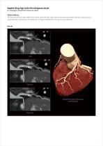
Napkin-Ring Sign in the first diagonal vessel Dr. Tatsugami, Hiroshima University, Japan Patient History This 60-year-old man with a BMI 29 and heart rate of 61 bpm had a history of acute myocardial infarction and a previous revascularization procedure. As a follow-up, CT was performed for coronary artery evaluation. Global Illumination Rendering with PIQE 1024
Open the catalog to page 7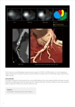
lumen low attenuation core non-calcified plaque high attenuation peripheral rim Global Illumination Rendering with PIQE 1024 The D1 shows a non-calcified plaque causing a stenosis scored as CAD-RADs 3 (50-69% stenosis). It is very challenging to confirm whether there is a napkin-ring sign in the DLR and PIQE 512 images. However, the PIQE 1024 images clearly shows a napkin-ring sign. Clinical Benefit The unique combination of high resolution, low contrast detectability and low noise enabled by PIQE 1024 makes it possible to identify vulnerable plaques with the napkin-ring sign. Therefore, PIQE...
Open the catalog to page 8All Canon Medical Systems Europe catalogs and technical brochures
 Alphenix 4D CT
9 Pages
 Automation Platform
6 Pages
 Radrex
12 Pages
 Xantara
12 Pages
 Ultimax-i
16 Pages
 Aceso+
12 Pages
 Aceso
12 Pages
 Artemis
8 Pages
 Viamo sv7
12 Pages
 Viamo c100
12 Pages
 Xario 100 MX Edition
8 Pages
 Xario 200g
12 Pages
 Xario 100g
8 Pages
 Aplio a550
20 Pages
 Aplio a450
16 Pages
 Aplio a
24 Pages
 Aplio i900
20 Pages
 Aplio i800
20 Pages
 Aplio i700
12 Pages
 Aplio i600
12 Pages
 Aquilion Exceed LB
12 Pages
 Aquilion LB
9 Pages
 Aquilion ONE / GENESIS Edition
21 Pages
 Aquilion ONE / PRISM Edition
27 Pages
 Aquilion Lightning SP
24 Pages
 Aquilion Lightning
17 Pages
 Aquilion Prime SP
21 Pages
 Aquilion Precision
19 Pages
 Aquilion Start brochure
21 Pages

- Ultrasound system
- Analysis software
- B/W ultrasound system
- Color doppler ultrasound system
- Portable ultrasound system
- Multipurpose ultrasound imaging system
- Radiology software
- Visualization software
- Tablet computer software
- Tablet PC software
- Digital radiography system
- Convex-array ultrasound system
- Linear-array ultrasound system
- Multipurpose radiography system
- X-ray system
- On-platform ultrasound system
- Multipurpose radiography X-ray system
- Diagnostic software
- Fixed ophthalmic examination
- Treatment software