Image-Guided Abdominal Biopsy Phantom

Image-Guided Abdominal Biopsy Phantom
The Model 071A Image-Guided Abdominal Biopsy Phantom is designed for training and demonstrating image-guided needle biopsy procedures. It is constructed from a self-healing material called Zerdine, allowing for multiple biopsy insertions with minimal needle tracking.
Features
- Contains 11 randomly positioned lesions (8-12 mm) and a 25 mm lesion near the vertebrae.
- Simulated spine and ribs are included.
- Visible under ultrasound, CT, and MRI.
- Anechoic solid polymer gel background that does not leak when punctured.
Benefits
- Improves performance of freehand abdominal biopsies.
- Tests new equipment and validates automated biopsy systems.
- Suitable for CT, MRI, and Ultrasound.
Specifications
- Dimensions: 28 cm x 20 cm x 12.5 cm (11" x 7.9" x 4.9").
- Weight: 5.5 kg (12.1 lb).
- Materials: Zerdine for background and masses, epoxy for ribs and vertebrae, urethane for scan surface.
Optional Features
- CT DICOM Data Set with 1.5 mm slice thickness at 120 kvp.
Additional Information
The phantom includes a foam-lined hard carry case and offers value-added services such as phantom-specific CMM, reference CT or MRI data sets, and customer-specific registration devices. To extend the phantom's lifetime, it is recommended to use higher gauge needles that are wetted and de-aired before insertion.
Certification
Certified by UL DQS Inc. to ISO 9001:2008, Certificate Registration No.10000905-QM08.
Catalog excerpts

Image-Guided Abdominal Biopsy Phantom Model 071A CT/ ULTRASOUND/ MRI IMAGE FUSION • LIVE SCANNING• BIOPSY TRAINING Visualize Biopsy Insertions with Minimal Needle Tracking The Model 071A, Image-Guided Abdominal Biopsy Phantom is a simplified abdominal phantom suitable for training and demonstrating image-guided needle biopsy navigation tools or procedures that require a constant visual reference for needle placement. Because it is constructed of a self-healing formulation of Zerdine, the phantom allows multiple biopsy insertions with minimal needle tracking. commodate image fusion techniques, CIRS can offer value added services such as phantom specific CMM, reference CT or MRI data sets, attachment of customer specific registration devices and inclusion of special point markers. The phantom contains 11 randomly positioned lesions, with sizes ranging from 8 to 12 mm. It also includes simulated spine, ribs, and a 25 mm lesion near the vertebrae. • mprove performance of freehand abdominal biopsies I • Validate automated biopsy systems The lesions and spine are visible under ultrasound, CT and MRI. The solid polymer gel background is anechoic and will not leak with punctured.* The phantom includes a foam lined hard carry case. To ac- *NOTE: Some permanent tracking may be evident if debris and air bubbles are entrained in the gel during the biopsy procedure. To extend the lifetime of the phantom, the use of higher gauge needles that have been wetted and de-aired prior to insertion is recommended. 2428 Almeda Avenue Suite 316 • Norfolk, Virginia 23513 • USA Tel: 800.617.1177 • 757.855.2765 • Fax: 757.857.0523 WWW.CIRSINC.COM Tissue Simulation & P
Open the catalog to page 1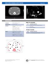
IMAGE-GUIDED ABDOMINAL BIOPSY PHANTOM SPECIFICATIONS DIMENSIONS PHANTOM WEIGHT COMPONENT DESCRIPTION Image-Guided Abdominal Biopsy Phantom User Guide Ribs & Vertebrae: Epoxy Scan Surface: Urethane OPTIONAL FEATURES Masses: Zerdine, soft tissue MASSES Large Mass: 25 mm Diameter Qty: 1 Positioned near vertebrae COMPONENT DESCRIPTION CT DICOM Data Set (Serial number specific, 1.5 mm slice thickness @ 120 kvp) Other Masses: 8-12 mm Diameter Qty:11 Randomly positioned in Background 2013 Computer Imaging Reference Systems, Inc. All rights reserved. Specifications subject to change without notice. Publication:...
Open the catalog to page 2All CIRS catalogs and technical brochures
 Model 002LFC
2 Pages
 Breast Elastography Phantom
2 Pages
 Prostate Training Phantom
2 Pages
 MRI-LINAC Dynamic Phantom
8 Pages
 UltraiQ
2 Pages
 E2E SBRT Phantom
8 Pages
 Model 711-HN
2 Pages
 Model 701-706
16 Pages
 Model 062M, 062MA, 062MQA
16 Pages
 Model 602
2 Pages
 Model 004
16 Pages


- Test phantom
- Tomography test phantom
- Radiography test phantom
- CT scan test phantom
- General purpose test phantom
- Ultrasound imaging test phantom
- Torso test phantom
- MRI test phantom
- Head test phantom
- Radiation therapy test phantom
- Breast test phantom
- Pediatric test phantom
- Abdomen test phantom
- Mammography test phantom
- Pelvis test phantom
- Whole body test phantom
- Skull test phantom
- Lung test phantom
- Fluoroscopy test phantom
- Brain test phantom