Model 004

Model 004
The CIRS Model 004 CT Simulator is designed for bone mineral analysis, focusing on the lumbar spine to assess trabecular bone density. It standardizes bone mineral content (BMC) measurement using CT technology, addressing variability issues across different scanners and patient anatomies.
Quantitative CT (QCT)
QCT quantifies human tissue characteristics using a CT scanner and a reference standard. Initially developed for lung nodule calcium evaluation, it now assesses bone mineral density, especially in the lumbar spine.
CT and Patient Variability
CT numbers vary due to scanner type, software, exposure factors, and patient anatomy. The CIRS Model 004 provides a machine and patient-independent reference standard.
The CIRS Approach
The Model 004 simulates average patient anatomy with materials equivalent to human tissues, accounting for age-related variations, ensuring consistent and accurate density measurements across CT scanners.
Features
- Simulates human tissue size, shape, and density.
- Includes vertebral inserts of varying density for accurate correlation.
- Accounts for age-related changes in marrow fat and mineral content.
- Compatible with all CT scanners, requiring no special software.
- Includes PC-based report software for data analysis.
Comparison with Other BMD Measurements
The CIRS system uses calcium hydroxyapatite (HA) for BMC values, offering more accurate representation compared to systems using dipotassium phosphate (K2HPO4). It corrects for marrow fat buildup affecting BMC measurements.
Normal Values and Comparisons
BMD values are compared with age- and sex-matched controls, expressed as Z-scores and T-scores, aiding in assessing bone density relative to a normal population.
Conclusion
The CIRS Model 004 CT Simulator provides a reliable and standardized method for bone mineral analysis, addressing variability issues and offering accurate bone health assessments.
- Measurement Sensitivity: Sensitivity of bone loss or gain measurement depends on precision and expected changes. DXA of the spine has higher diagnostic sensitivity compared to SPA of the forearm.
- Slice Thickness Qualification: Use 5 mm or 10 mm slice sections for vertebral BMC analysis. Ensure slice thickness accuracy to avoid including cortical bone.
- Beam Evaluation: Evaluate images based on slice thickness and rotation. Proper alignment is crucial for accurate measurements.
- Reconstruction Algorithms: Use body imaging algorithms recommended by the scanner manufacturer. Avoid algorithms for targeted reconstructions.
- Beam Pulse and Scan Time: Use manufacturer-recommended kVp and ensure adequate exposure. Avoid streak artifacts for reliable analysis.
- Circle of Reconstruction: Use the largest circle of reconstruction possible to avoid incorrect values due to patient overlap.
Marrow Fat Correction
Bone marrow fat increases with age, affecting CT value readings. A correction method ensures accurate bone mineral content measurements.
Scanning and Data Acquisition Procedure
- Position the patient and obtain a lateral scout view to localize the lumbar area.
- Scan the lumbar area using the largest circle of reconstruction possible.
- Set up the simulator with appropriate vertebral inserts and scan using the same technique as for the patient.
- Use CIRSCALC software for data analysis and interpretation.
CIRSCALC Software
The CIRSCALC software is used for data analysis and report generation, featuring patient record management, exam data updates, and patient history maintenance.
Conclusion
This document provides comprehensive guidelines for accurate bone mineral content measurement using CT analysis, emphasizing proper technique, alignment, and marrow fat correction.
Deleting a Patient: Navigate to Patient Exams under the File Menu, select the patient using the Binocular look-up, double-click the patient, click the Red “X” button to delete, verify the deletion, and exit.
Update Marrow Fat Correction: Access System Configuration from the File menu, make changes using the Edit Icon, save, and exit.
Data Back-Up: Use the Standard Windows Back-up function located in Start/Programs/Accessories/System Tools.
Bone Mineral Content (BMC) Calculation: CIRSCALC software calculates vertebral BMC using vertebral ROI values and simulator ROI values. Bone mineral values differ by 20 mg/cc from other systems due to calcium hydroxyapatite use.
Printing Reports: Select the Patient Folder, choose the patient, and select Reports from the toolbar to print the BMC Bone Densitometry Graph.
Building Normal Value Table: Create a Normal Value Table by selecting the file from the toolbar, adding a new table, building it, saving, and exiting.
Reviewing Images: Radiologists should review images for technique adherence and additional findings. Quantitative CT assists but does not replace physician judgment.
Medicare and Reimbursement Guidelines: Proper ICD-9-CM codes must accompany CPT codes for Medicare claims. Coverage varies by payer and locality. CPT 77078 is used for CT Bone Densitometry.
Product Warranty and Returns: The CIRS Model 004 has a 48-month warranty. Unauthorized modifications void warranties. For returns, contact Customer Service for an RMA number and follow return instructions.
References: The document includes references to studies and publications related to bone densitometry and osteoporosis.
Catalog excerpts
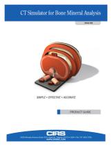
CT Simulator for Bone Mineral Analysis Model 004 SIMPLE • EFFECTIVE • ACCURATE PRODUCT GUIDE 2428 Almeda Avenue Suite 316 • Norfolk, Virginia 23513 • USA • Tel: 757-855-2765 WWW.CIRSINC.COM
Open the catalog to page 1
Background Since the advent of whole-body Computed Tomography (CT) scanners in the late 1970’s, considerable research effort has been expended to develop a method of density measurement. The power of the CT scanner could be applied to studies of bone demineralization, particularly in the lumbar region of the vertebral column where the earliest changes of osteoporosis or diffuse bone loss occur. It became apparent to early researchers that several problems of scanner variability needed to be overcome before quantitative studies could be generally performed with high reliability. Quantitative CT...
Open the catalog to page 2
The CIRS Approach to Quantitative CT PRINCIPLES OF DESIGN The CIRS CT Model 004 Simulator for Bone Mineral Analysis is designed to take into account all the known sources of variance affecting the measurement of density in the vertebral area by simulating the average patient’s anatomy in terms of shape and density by using materials essentially equivalent to human tissues as far as X-ray interactions are concerned, including age-related variations in vertebral composition. • Accurately simulates the size, shape and CT density of human tissue The design of the system permits reduction of all sources...
Open the catalog to page 3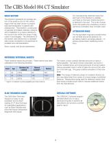
The CIRS Model 004 CT Simulator BASIC SECTION Our manufacturing methods insure that each part of the phantom is carefully controlled so that each finished phantom is identical to the next. This is why human bone, the quality and consistency of which cannot be controlled, have not been used. The phantom represents an average section of the anatomy (2nd to 4th lumbar region) that has been shown to present early demineralization in patients with osteoporosis. The phantom, constructed of specially formulated materials, interacts with X-Radiation in a manner identical to the human body (within the...
Open the catalog to page 4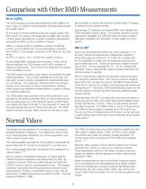
Comparison with Other BMD Measurements HA vs. K2HPO4 late the effect of marrow fat build-up in performing CT analysis of vertebral bone mineral content. The CIRS simulator provides Bone Mineral Content (BMC) values in mg/cc of calcium hydroxyapatites (HA) that approximate ash weight values. Dual energy CT evaluation of vertebral BMC has validated the CIRS simulation system values. For scanner operators having dual energy capability, the CIRS Simulator provides a system calibration capability, but simulation of each patient is not required. At the range of normal vertebral body ash weight values...
Open the catalog to page 5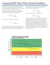
Comparing BMD Values With a Normal Population The BMD measured for a patient is compared with age- and sex- matched controls as well as with sex-matched young healthy controls. The values are then expressed as percentiles or standard deviation scores, called Z- or T-scores. The T-score is a measure of the difference between the patient’s BMD and the mean BMD of young normals. The Tscore, in SD values, is calculated as: The Z-score is a measure of the difference between the patient’s BMD and the mean BMD of age- and sex-matched peers. The Z-score, in SD values, is calculated as: Z-score(SD) =...
Open the catalog to page 6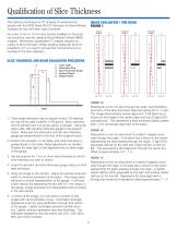
Qualification of Slice Thickness IMAGE EVALUATION 1 MM BEAM FIGURE 1 The optimum technique for CT analysis of vertebral bone density with the CIRS Mode 004 CT Simulator for Bone Mineral Analysis will vary with each type of scanner. As a rule, 5 mm or 10 mm slice section available on the scanner should be used for vertebral Bone Mineral Content (BMC) analysis. Remember, quantitative CT analysis requires accuracy of slice thickness. (When isolating trabecular bone for analysis by CT, you want to ensure that cortical bone is not included in the slice selected.) SLICE THICKNESS AND BEAM EVALUATION...
Open the catalog to page 7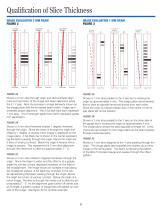
Qualification of Slice Thickness IMAGE EVALUATION 3 MM BEAM FIGURE 2 0.0 deg Rotation Beam Profile Image Plane Beam Profile Image Plane IMAGE EVALUATION 1 MM BEAM FIGURE 3 2.0 deg-CW Rotation Beam Profile Image Plane Beam Profile FIGURE 2A Shows a 3 mm slice through origin and demonstrates alignment and symmetry of the image and beam alignment along the X / Y axis. Note the increase in image elements shown on the image plane with the increased beam width. Image demonstrates proper alignment. The 3 full dark bars each represent 1 mm slice. The 2 full length lighter bars (20%) represent partial...
Open the catalog to page 8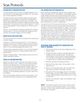
Scan Protocols ALGORITHM OF RECONSTRUCTION On some scanners there is only one available algorithm of reconstruction for body applications and there is no other option but to use the algorithm provided. Bone marrow fat increases with age, and this increased bone marrow fat affects the CT value reading. Theoretically, a person could maintain the same level of bone mineral content over a period of years and yet have progressively lower CT values due solely to the increase in bone marrow fat and the way in which this increase affects the machine’s reading. This effect has been described by a number...
Open the catalog to page 9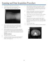
Scanning and Data Acquisition Procedure 1. Place patient on table with arms over head. Obtain a lateral scout view (scanography or pilot view) to localize third lumbar vertebral area. Set up simulator on support board, properly sized and with vertebral insert for 50 mg/cc (age 45) of mineral content in place (be sure to attach velcro backed “mark- er” to support board so that retained images will reflect which insert was used). Position with laser light in the center of the simulator. (Ensure the simulator is vertical to the table use a sandbag on the foot of the stand if necessary). Scan simulator...
Open the catalog to page 10
CIRSCALC® Software CIRSCALC is PC software for use with the CIRS Model 004 Lumbar Reference Simulator. The program performs data analysis calculations and produces printed reports. ADDING A NEW PATIENT FOLDER 1. Select the Patient’s Exam Folder by selecting Patient Exam under the File Menu. The CD contains both “New Installation” and “update” for previous users of Version 6.01. For questions pertaining to this program or the Bone Mineral Measurement System, or to obtain the latest version of CIRSCALC, please contact CIRS. 2. Click the ADD button (hint: move the mouse pointer slowly over the buttons...
Open the catalog to page 11All CIRS catalogs and technical brochures
 Model 002LFC
2 Pages
 Breast Elastography Phantom
2 Pages
 Prostate Training Phantom
2 Pages
 MRI-LINAC Dynamic Phantom
8 Pages
 UltraiQ
2 Pages
 E2E SBRT Phantom
8 Pages
 Model 711-HN
2 Pages
 Model 701-706
16 Pages
 Model 062M, 062MA, 062MQA
16 Pages
 Model 602
2 Pages


- Test phantom
- Tomography test phantom
- Radiography test phantom
- CT scan test phantom
- General purpose test phantom
- Ultrasound imaging test phantom
- Torso test phantom
- MRI test phantom
- Head test phantom
- Radiation therapy test phantom
- Breast test phantom
- Pediatric test phantom
- Abdomen test phantom
- Mammography test phantom
- Pelvis test phantom
- Whole body test phantom
- Skull test phantom
- Lung test phantom
- Fluoroscopy test phantom
- Brain test phantom