CT Protocol Guide

CT Protocol Guide
Catalog excerpts
CT Protocol Reference Guide For the ConforMIS Family of Knee Resurfacing Implants
Open the catalog to page 1
PATIENT POSITION IMAGE AQUISITION —Protocol Chart IMAGE ARCHIVING IMAGE DATA TRANSFER All questions regarding this protocol reference guide should be addressed to: ConforMIS Imaging Support 28 Crosby Drive Bedford, MA 01730-9998 Tel: 781/345-9170 Email: [email protected] ConforMIS Imaging Support is available: Monday-Friday 8.00 am – 6.00 pm (Eastern Time)
Open the catalog to page 2
Patient Position: The patient should be at isocenter in the gantry and must be supine with extremity of interest fully extended. To ensure our ability to correct for malalignment of the knee it is critical that the foot be perpendicular to the table with toes pointing straight up and secured to prevent motion. Do not place a sponge or pillow beneath the knee or ankle. *** When an implant or other device is present in the opposite knee, please make every effort to position that knee flexed and out of the FOV to reduce the artifacts in the affected knee joint. Please use a metal artifact reduction...
Open the catalog to page 3
ConforMIS CT Scan Protocol Series 1 FULL LEG, Hip through Ankle Kernel / Algorithm Hip – Femoral head only (acetabulum only) Knee – distal 1/3 of the femur through proximal 1/3 of tibia (should include the entire patella through the fibula head) Ankle – center at tibiotalar joint space scan 2cm above the joint to 2cm below Axial Reconstruction Thickness X Increment Multi Planar Reformat– knee only Multi Planar Reformat– knee only
Open the catalog to page 4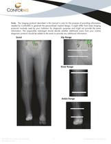
Note: The imaging protocol described in this manual is only for the purpose of providing information needed by ConforMIS to generate the personalized implant design. It might differ from knee imaging protocols routinely used by your institution for diagnostic purposes and might not provide the same information. The responsible radiologist should decide whether additional scans from your routine diagnostic protocol should be added to the exam to provide any additional information.
Open the catalog to page 5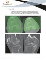
Knee MPR: Sagittal and Coronal reformats should be limited to the side of interest and cover bone to bone only. It is preferred for Coronal MPR to be parallel to femoral condyles, and Sagittal MPR to be perpendicular to femoral condyles.
Open the catalog to page 6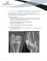
CT Arthrography Protocol: OPTIONAL and is NOT required for a ConforMIS protocol scan This CT imaging option is most often used to assist in the diagnostic evaluation of the patellofemoral cartilage and the cartilage in the other tibiofemoral compartment. It can also be used for evaluating menisci and cruciate ligaments. 3.1 Routine Arthrography: A routine knee arthrogram should be performed using a contrast agent concentration of approximately 150 mg of iodine per milliliter. The dilution is important for the visualization of the bone and soft tissue structures in the joint space. Example 1:...
Open the catalog to page 7
Image Archive Important: Your site must keep a permanent archive (PACS) copy of the knee CT exams. Image Data Transfer: ***It is critical that ConforMIS protocol scans are sent immediately upon completion of the exam via electronic upload whenever possible to ensure the best possible care for the patient.*** There are several methods of image transfer available for ConforMIS protocol exams. 5.1 Image Transmission - Secure Web Upload: ConforMIS exams can be uploaded from a CD, DVD, or a web enabled PACS to our secure website. Go to http://www.ConforMIS.com/Imaging-Professionals/Upload-a-Scan to...
Open the catalog to page 8


