


Improving cell viability
Improving cell viability
LED illumination in widefield fluorescence microscopy offers significant advantages in minimizing photodamage, which includes phototoxicity and photobleaching, in both fixed and live cells. This document explores strategies to enhance cell viability and reduce photodamage using LED systems.
Photodamage can skew experimental results and is not always immediately apparent. Traditional lamps like mercury or metal halide can easily damage samples, whereas LEDs offer enhanced control to mitigate these effects. Phototoxicity affects live cell imaging by generating reactive oxygen species (ROS) that damage cellular components. Photobleaching affects both fixed and live samples by degrading fluorophores, leading to data misinterpretation.
- Gentle Illumination: Use the lowest possible light levels to observe samples. LED systems allow precise control of irradiance, unlike traditional lamps that require neutral density filters.
- Exposure Time and Irradiance Balance: Lowering irradiance while increasing exposure time can reduce photodamage, allowing cellular detoxification systems to manage ROS.
- Reduce Illumination Overhead: LED systems with electronic control reduce unnecessary exposure by synchronizing with cameras, minimizing the time samples are exposed without data collection.
- Lower Energy Wavelengths: LEDs offer a range of wavelengths, including lower energy options that are less damaging to samples.
- Strobe Illumination: Using strobe lighting instead of constant illumination can significantly reduce photobleaching by allowing triplet states to reset.
Modern LED systems provide sufficient light for fluorescence microscopy while offering control features to minimize photodamage. Optimizing illumination settings and exploring new fluorophore and filter combinations are recommended for effective widefield fluorescence experiments.
The document includes references to studies and articles for further exploration of photodamage and LED illumination strategies.
Catalog excerpts
Improving cell viability Minimising photodamage during fluorescence microscopy with LED illumination Amongst the many benefits of LED illumination for widefield fluorescence microscopy is the potential for minimising the serious problem of photodamage in both fixed and live cells. Whether running a complete experiment or simply finding the region of interest prior to applying confocal microscopy, LED illumination systems are well suited to minimise both types of photodamage: phototoxicity and photobleaching. Find out: • Photodamage is not always obvious but can still skew results • Mercury or metal halide lamps can easily damage samples • LEDs reduce photodamage via enhanced control and TTL camera synchronisation • Controlling widefield illumination is also important when setting up confocal experiments Simply Better Control
Open the catalog to page 1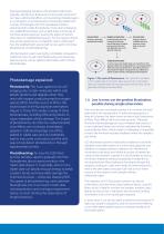
Exposing biological samples to the excitation light levels typically used during a fluorescence microscopy experiment can have a detrimental effect, and countering photodamage is a crucial aspect of any fluorescence microscopy experiment.1 A variety of strategies exist, from specialised media to selecting bright, stable fluorophores.2 Even when employing non-widefield techniques such as light sheet microscopy to minimise sample exposure, locating the region of interest often relies on widefield fluorescence. For the majority of light microscope experiments, it is therefore vital to understand...
Open the catalog to page 2
@LaissuePhilippe Hey #microscopytwitter, many thanks for all your answers re #happycells! Here's our last poll (for now) on #phototoxicity in #microscopy: If your live experiment seems to be affected by phototoxicity, what do you do? I=intensity (of excitation light), t=exposure time (of camera) Reduce I, increase t 65.2% Use better 'scope / label 25.9% 112 votes • Final results 12:20 PM Oct 15, 2020 © C? 5 Q 10 <9 Copy link to Tweet One proposed explanation is that by exposing the sample to a lower number of photons at any one time point, the cell's detoxification systems can cope with clearing...
Open the catalog to page 3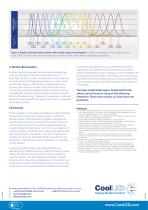
Figure 4: Modern LED illumination systems offer a wide range of wavelengths. Available wavelengths for the single-channel CoolLED pE-100 allow the use of wavelengths into the near-IR, which are less damaging to samples. Another interesting approach to reducing photobleaching is the use of strobe instead of constant illumination.2, 12, 13 Essentially, the theory is that constant illumination continues to create atoms in the fluorophore which exist in the triplet excited state (Figure 1). When light is instead delivered as a strobe with microsecond gaps, these triplet states have time to reset...
Open the catalog to page 4All CoolLED catalogs and technical brochures
 pE-400 Series
4 Pages
 pT-100
2 Pages

