aview: Lung Texture
1 /8Pages

aview: Lung Texture
1 /8Pages
Catalog excerpts

Lung Texture Full-automatic lung texture pattern analysis
Open the catalog to page 1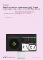
Deep learning AI technology automatically detects and analyzes major patterns of interstitial lung disease. core:line's aview:Lung Texture defines the fibrotic area of the lungs in 6 patterns and automatically generates a report by analyzing the location and distribution. Quantitative analysis and results aids, the difficult reading cases and reduces deviations due to the reading technique. Lung Texture
Open the catalog to page 2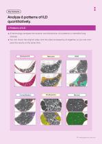
Analyze 6 patterns of ILD quantitatively. 6 Patterns of ILD AI technology analyzes the location and distribution of 6 patterns in interstitial lung disease. You can check the original video and the video analyzed by AI together, so you can compare the results at the same time.
Open the catalog to page 3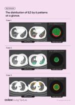
The distribution of ILD by 6 patterns at a glance. Case 1. Lung Texture
Open the catalog to page 4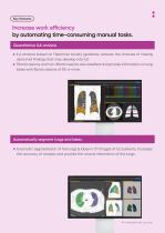
Increase work efficiency by automating time-consuming manual tasks. Quantitative ILA analysis ILA analysis based on Fleischner society guidance, reduces the chances of missing abnormal findings that may develop into ILD. Fibrotic lesions and non-fibrotic lesions are classified and provide information on lung lobes with fibrotic lesions of 5% or more. Automatically segment lungs and lobes. Automatic segmentation of the lungs & lobes in CT images of ILD patients, increases the accuracy of analysis and provide the volume information of the lungs.
Open the catalog to page 5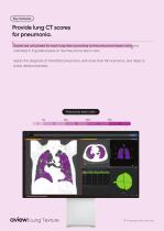
Provide lung CT scores for pneumonia. Scores are calculated for each lung lobe according to the pneumonia lesion ratio and indicated in 5 grades based on the Pneumonia lesion ratio. Assists the diagnosis of interstitial pneumonia, with more than 150 variations, and helps to probe related diseases. Pneumonia lesion ratio 5% 1 Lung Texture
Open the catalog to page 6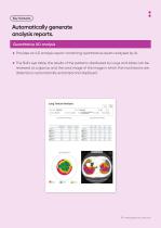
Automatically generate analysis reports. Quantitative ILD analysis Provides an ILD analysis report containing quantitative results analyzed by AI. The Bull’s eye table, the results of the patterns distributed by lungs and lobes can be reviewed at a glance, and the axial image of the image in which the most lesions are detected is automatically extracted and displayed.
Open the catalog to page 7
Lung Texture Full-automatic ILD texture pattern analysis ILD / ILA / Pneumonia Prosper with Better Health
Open the catalog to page 8All Coreline catalogs and technical brochures
 aview: Research
6 Pages
 aview: RT ACS
4 Pages
 aview: CAC
8 Pages
 aview:LCS
8 Pages
- Analysis software
- Visualization software
- Radiology software
- Tablet computer software
- Tablet PC software
- Reporting software
- Diagnostic software
- Planning software
- Cloud-based software
- Automated software
- Treatment software
- Traceability software
- Data management software
- AI-assisted software
- Artificial intelligence software
- Web-based software
- Printing software
- Test software
- Server software
- CT software