- Catalogs
- Cortex-Dental Implants Industries
- Synthe-Bone
- Products
- Catalogs
- News & Trends
- Exhibitions
Synthe-Bone

Synthe-Bone
Synthe-Bone is a range of bone replacement products made from Tricalcium Phosphate, adhering to ASTM F1088-04 standards. It is designed for odontological use, mimicking the structure of spongy bone to support osteoconduction and facilitate bone regeneration.
Specifications and Composition
Synthe-Bone features interconnected porosity similar to spongy bone, promoting osteointegration and bioreabsorption. It is bioactive, allowing full integration and replacement by the patient's own bone. The product is composed of Beta-Tricalcium Phosphate with a purity greater than 99%.
Characteristics and Benefits
- Biofunctionality and Osteoconduction: Provides mechanical stability and preserves defect shape and volume.
- Macroporosity and Microporosity: Enhances cell adhesion and growth factor integration, supporting bone regeneration.
- Bioabsorption and Bioremodelling: Reacts with physiological mediums to form hydroxyapatite, leading to new bone formation.
Regenerative Process
Over a period of 3 weeks to 14 months, Synthe-Bone supports predictable and stable bone regeneration, as evidenced by micrographs showing the progression from initial cell colonization to mature bone formation.
Indications for Use
- Filling post-extraction bone cavities
- Crest reconstruction
- Covering fenestrations
- Intrabone defects in periodontics
- Expanding bone regeneration
- Furcal lesions and radicular exposures
Frequently Asked Questions
- Use with Antibiotics: Generally unnecessary; mixing with antibiotics is not recommended.
- Mixing with Autologous Bone: Can be mixed to increase implant volume, though designed for standalone use.
- Re-sterilization: Not authorized for re-sterilization in dental practice.
Manufacturer's Note
KERAMAT, the manufacturer, disclaims responsibility for unauthorized re-sterilization.
Catalog excerpts
Reasorbable Bone Regenerator The Future of Dental Implants
Open the catalog to page 1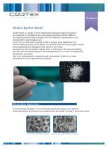
What is Synthe-Bone? Synthe-Bone is a range of bone replacement products made of Tricalcium (3-Phosphate in compliance with international standard ASTM F1088-04. The KeraOs product range complies with the maximum requirements of any biomaterial for odontological use. Synthe-Bone’s structure is similar to that of spongy bone trabecules in its interconnected porosity, which allows it to work as osteoconductor support where blood capillaries and osteogenic cells adhere t form bone. Its bioactivity and composition allows them to intervene in the bone remodeling process with full oesteointegration...
Open the catalog to page 2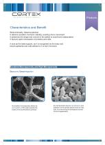
The Future of Dental Implants Biofunctionality. Osteoconduction. It delivers excellent mechanic stability, avoiding micro-movement. It preserves the shape and volume of the defect to avoid bone reabsorption. It favours rapid colonisation of proteins and cells. It acts as the ideal support, as it is recognised by the body and blood capillaries and cells adhere to it to form the bone. Excellent Macroporosity and High Microporosity Bioactivity Osteointegration Its excellent microporosity allows for Its microporosity favours cell adhesion and permeability of the cells towards the adhesion of the...
Open the catalog to page 3
Beta-Tricalcium Phosphate Purity > 99% Bioabsorption Bioremodelling • Synthe-Bone superficially reacts with its physiological medium, dissolving and precipitating hydroxyapatite on the surface. • This precipitation leads to the appearance of osteoblasts and collagen fibre that will form immature bone. • Immature bone becomes structured and mature, continuing with the absorption of Synthe-Bone until it is totally replaced by the newly formed bone. Predictable and stable regenerative results “Effective Bone Regeneration” (a) Goldner staining (b) (c) Wheatley staining
Open the catalog to page 4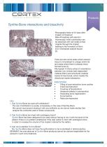
Synthe-Bone interactions and bioactivity Micrographs taken at 45 days after implant of Tricalcium Beta-Phosphate with electron microscopy. Initial colonization can be observed with cell nodes (I) that migrate through the implant leading to the formation of fibrin (non mineralized osteoid tissue) (2). There are also some areas where osteoid tissue is mineralized to a larger extent (3) and areas where there is already newly formed bone (4). Micrograph A shows areas of coexistence between non mineral-ized reabsorbed material (fibrin) and structured material (newly formed bone), which implies the...
Open the catalog to page 5
The Future of Dental Implants
Open the catalog to page 6All Cortex-Dental Implants Industries catalogs and technical brochures
 Product Catalogue 2015
25 Pages
 Magix
Magix2 Pages
 Product Classification
1 Page
 Surgical Manual
37 Pages
 DRILL STOPPER KIT
1 Page


Archived catalogs
 NON-TOUCH PACKAGE
1 Page
- Implant abutment
- Titanium implant abutment
- Straight implant abutment
- Dental surgery instrument kit
- Titanium dental implant
- Internal implant abutment
- Conical dental implant
- Angled implant abutment
- Straight dental implant
- Dental implant surgery instrument kit
- Drill bit
- Hexagonal implant abutment
- Screw implant abutment
- Internal hexagon implant abutment
- Dental implant analog
- External implant abutment
- Hexagonal dental implant
- Dental drill bit
- Multi-unit implant abutment