- Catalogs
- CyberLogic
- ULTRASONIC ASSESSMENT OF THE RADIUS IN VITRO
ULTRASONIC ASSESSMENT OF THE RADIUS IN VITRO
1 /8Pages
ULTRASONIC ASSESSMENT OF THE RADIUS IN VITRO
1 /8Pages
Catalog excerpts
Ultrasound in Med. & Biol., Vol. 34, No. 12, pp. 1972–1979, 2008 Copyright © 2008 World Federation for Ultrasound in Medicine & Biology Printed in the USA. All rights reserved 0301-5629/08/$–see front matter ● Original Contribution ULTRASONIC ASSESSMENT OF THE RADIUS IN VITRO VINCENT LE FLOCH,*† GANGMING LUO,†‡§ JONATHAN J. KAUFMAN,†¶ and ROBERT S. SIFFERT¶ *Ecole Nationale Superieure d’Arts et Metiers, Aix-en-Provence, Provence-Alpes-Cote-d’Azur, France; CyberLogic, Inc., New York, NY, USA; ‡VA New York Harbor HealthCare System, New York, NY, USA; §New York University School of Medicine, Dept of Rehabilitation Medicine; and ¶Department of Orthopedics, The Mount Sinai School of Medicine, New York, NY, USA † (Received 23 January 2008; revised 15 May 2008; in final form 22 May 2008) Abstract—The overall objective of this research is to develop an ultrasonic system for noninvasive assessment of the distal radius. The specific objective of this study was to examine the relationship between geometrical features of cortical bone and ultrasound measurements in vitro. Nineteen radii were measured in through transmission in a water bath. A 3.5 MHz rectangular (1 cm x 4.8 cm) single element transducer served as the source and a 3.5 MHz rectangular (1 cm x 4.8 cm) linear array transducer served as the receiver. The linear array consisted of 64 elements with a pitch of 0.75 mm. Ultrasound measurements were carried out at a location that was 1/3rdrd of the length from the distal end of each radius and two net time delay parameters, NetDW and NetCW, associated with a direct wave (DW) and a circumferential wave (CW), respectively, were evaluated. The cortical thickness (CT), medullar thickness (MT) and cross-sectional area (CSA) of each radius was also evaluated based on a digital image of the cross-section at the 1/3rd location. The linear correlations between CT and NetDW was r ؍ 0.91 (p < 0.001) and between MT and NetCW ؊ NetDW was r ( 36.0 ؍p < 0.05). The linear correlation between CSA and a nonlinear combination of the two net time delays, NetDW and NetCW, was r ( 59.0 ؍p < 0.001). The study shows that ultrasound measurements can be used to noninvasively assess cortical bone geometrical features in vitro as represented by cortical thickness, medullar thickness and cross-sectional area. (E-mail: [email protected]) © 2008 World Federation for Ultrasound in Medicine & Biology. Key Words: Osteoporosis, Ultrasound, Radius, Cortical thickness, Cross-sectional area, Net time delay. increase the incidence of hip and other fractures as well (Anonymous 2001). The primary method for diagnosing osteoporosis and associated fracture risk relies on bone densitometry to measure bone mass (Kaufman and Siffert 2001). The use of bone mass is based on the well-established thesis that bone strength is strongly related to the amount of bone material present and that a stronger bone in a given individual is associated generally with a lower fracture risk (Johnell et al. 2005). Radiological densitometry, which measures the (areal) bone mineral density (BMD) at a given site (e.g., hip, spine, forearm) is currently the accepted indicator of bone strength and fracture risk (Johnell et al. 2005; Blake and Fogelman 2003). Clinically, this is often done using dual energy X-ray absorptiometry (DXA), which measures the BMD in units of g/cm2 (Bonnick 2004). Notwithstanding the fact that X-ray methods are useful in assessing bone mass and fracture risk, osteoporosis remains one of the largest undiagnosed and un- Osteoporosis is a significant health problem affecting more than 20 million people in the United States and more than 200 million worldwide (Anonymous 2001). Osteoporosis is defined as the loss of bone mass with a concomitant disruption in microarchitecture, leading to an increased risk of fracture (Kanis 2002). The most common osteoporotic fractures occur at the wrist, spine and hip. Hip fractures have a particularly negative impact on morbidity. Approximately 50% of those individuals suffering a hip fracture never live independently again (Miller 1978). Currently, there are about 200,000 hip fractures yearly in the United States and approximately 1 million worldwide (Anonymous 2001; Melton 1988). The aging of the worldwide population is expected to Address correspondence to: Jonathan J. Kaufman, PhD, CyberLogic, Inc., 611 Broadway, Suite
Open the catalog to page 1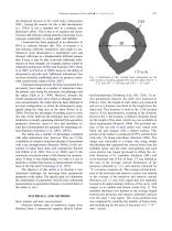
Ultrasonic assessment of the radius ● V. LE FLOCH et al. der-diagnosed diseases in the world today (Anonymous 2001). Among the reasons for this is that densitometry (i.e., DXA) is not a standard tool in a primary care physician’s office. This is due to its expense and inconvenience and reticence among patients concerning X-ray exposure, particularly in young adults and children. Ultrasound has been proposed as an alternative to DXA to estimate fracture risk. This is because it is non-ionizing, relatively inexpensive and simple to use. Moreover, since ultrasound is a mechanical wave and interacts...
Open the catalog to page 2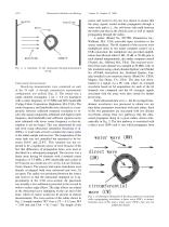
Ultrasound in Medicine and Biology Fig. 2. A schematic of the ultrasound through-transmission set-up. Ultrasound measurements Bench-top measurements were conducted on each of the 19 radii. A through transmission experimental configuration was utilized (Fig. 2). The source was a single element rectangular 1 cm ϫ 4.8 cm transducer with a center frequency of 3.5 MHz and 60% bandwidth (Valpey Fisher Corporation, Hopkinton, MA, USA). The center frequency and bandwidth were selected as a compromise between sufficient temporal resolution to resolve the signal components (obtained with higher center frequency...
Open the catalog to page 3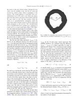
Ultrasonic assessment of the radius ● V. LE FLOCH et al. the source to the near cortical surface, through the near cortex, into the medullar cavity, into the far cortex and out the far cortical surface where it propagates to the receiver. The second pathway is associated with a circumferential wave (CW) and is one which also propagates from the source to the near cortical surface and into the near cortex as well, but then remains within the cortex as it “circumferentially” propagates within the bone cortex until it emerges at the far cortical surface and again propagates to the receiver. The...
Open the catalog to page 4Archived catalogs
 Ultrasound Simulation in Bone
14 Pages
 UltraScan™ 650
2 Pages