


EMG Sensor Placement

EMG Sensor Placement
This document provides guidance on the placement of Delsys Surface EMG Sensors, focusing on recording EMG data from forearm muscles. It highlights the importance of understanding both hardware and software for successful EMG recordings.
- The Surface EMG Signal: EMG signals are electrical activities from contracting muscles, detected through motor unit action potentials.
- Delsys EMG Sensors: These sensors use differential amplification to enhance signal quality by removing noise and amplifying desired signals. The design ensures consistent measurements.
- EMG Sensor Location and Placement: Proper placement is crucial for quality signal detection, avoiding edges, motor points, and tendons to minimize crosstalk.
- Experimental Set-up: Involves recording EMG data from forearm muscles using differential sensors to optimize signal detection.
- Test Configuration Design: Involves creating configurations for data acquisition using EMGWorks software.
- Data Acquisition: Data is collected with adjustments to sensor placement for optimal signal detection.
- Refinement of Sensor Location: Sensors are adjusted during contractions to ensure optimal signal detection.
- Align the sensor along muscle fibers, avoiding edges, motor points, or tendons.
- Use the sensor as a probe to find the optimal position during contractions.
- Prepare the skin and sensor surface with isopropyl alcohol and use adhesive interfaces for secure placement.
The experiment records EMG data from forearm muscles during wrist and finger movements. Proper sensor placement is critical due to muscle proximity, with adjustments made to maximize signals and minimize crosstalk.
Proper sensor placement is crucial for detecting quality EMG signals, with crosstalk minimized through careful sensor adjustment.
The document provides guidelines for EMG sensor placement, emphasizing anatomical knowledge and experimental design for accurate recordings.
The EMG sensor measures 41 x 20 x 5 mm, with a CMRR of -92 dB and noise level of 1.2 μV.
Includes a screenshot of recordings from three EMG sensors during muscle contractions, noting low amplitude activity from the right flexor carpi radialis.
Refer to the tutorial section of the knowledge center at www.delsys.com for proper EMG sensor placement.
Catalog excerpts

Technical Note 101: EMG Sensor Placement Purpose This technical note addresses proper placement technique for Delsys Surface EMG Sensors. The technique is demonstrated through an experiment in which EMG data is recorded from several forearm muscles. Hardware Concepts • • • • The Surface EMG Signal Delsys EMG Sensors EMG Sensor Location and Placement Experimental Set-up Software Concepts • Test Configuration Design • Data Acquisition • Refinement of Sensor Location www.delsys.com
Open the catalog to page 1
Introduction The key to acheiveing a successful EMG recording resides in understanding how to use and where to place the EMG sensors. This document presents important concepts in this regard, that relate to both Delsys® hardware and software. These concepts are presented from the vantage point of an experimental protocol and the ensuing analysis of the data. The intention of this structure, which is consistent throughout our Technical Notes Series, is to provide guidelines for working with Delsys products, and to promote creativity and sound judgement during the experimental design phase. The...
Open the catalog to page 2
EMG Sensor Location Proper EMG sensor location is critical for detecting quality surface EMG signals. The user should consult an anatomical atlas to determine the precise location, origin, insertion, and function of the muscle being studied as well as any nearby muscles that may produce underiable signals (crosstalk). 1. Place the sensor along the longitudinal midline of the desired muscle with the arrow parallel to the muscle fibers. DO NOT place the sensor at the outside edges of the muscle. In this region, the sensor is susceptible to detecting crosstalk signals from adjacent muscles. Place...
Open the catalog to page 3
EMG Sensor Placement Although the Delsys Surface EMG Sensor can be used as a probe without skin preparation or adhesive, the following simple steps are needed to assure optimal signal detection once the final location is determined. 1. Wipe the surface of the sensor and the silver detection bars with an isopropyl alcohol pad to remove residue. Allow the sensor to air dry for a few seconds. Shave excessive hair from skin at the detection site. Wipe the skin at the site with an isopropyl alcohol pad to remove oils and surface residues. Allow the skin to air dry for a few seconds. Apply the sensor...
Open the catalog to page 4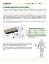
EMG Sensor Experiment - Hardware Setup The goal of this experiment is to record three channels of surface EMG data from a subject while performing different wrist and finger movements. The EMG data will be recorded from the right flexor carpi radialis, right flexor digitorum superficialis, and the right extensor carpi radialis longus. Since these muscles are all in close proximity, a thorough understanding of their anatomy and function is necessary, and proper EMG sensor location and placement is critical. In the experiment, each muscle will be contracted individually with Delsys Single Differential...
Open the catalog to page 5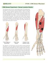
EMG Sensor Experiment - Sensor Location Details The illustrations below show the ventral (palmar) aspect of the right forearm. Each depicts one of the muscles of interest including its tendons of origin and insertion. The flexor digitorum superficialis shares its origin on the radius, ulna, and the distal humerus. Its tendons insert on the proximal aspect of each of the middle phalanges of the fingers. The flexor carpi radialis originates from the medial epicondyle of the humerus. It inserts at the proximal aspect of the second and third metacarpals. The extensor carpi radialis longus originates...
Open the catalog to page 6
EMG Sensor Experiment - Software Setup EMGWorks Acquisition requires the user to specify a Hardware Configuration and a Test Configuration to use for recording data. Please consult the EMGWorks manual for more details on the procedure for creating these. Hardware Configuration A Hardware Configuration should be created according to the hardware that you are using. In this case, the configuration was created for a Bagnoli 16-Channel EMG System and a National Instruments M-Series A/D Card. Test Configuration A Test Configuration should be created consisting of 6 Sets. The Test Control window in...
Open the catalog to page 7
Data Analysis The screenshot below shows the recordings from all three EMG sensors during contractions performed by the right flexor carpi radialis. It can be seen that the surface EMG activity from the right flexor carpi radialis is obviously of the greatest magnitude. There is, however, low amplitude surface EMG activity detected by the sensors over the right flexor digitorum superficialis and the right extensor carpi radialis longus as well. This detected activity was minimized by adjusting the locations of the sensor. This activity may be crosstalk from the right flexor carpi radialis. Many...
Open the catalog to page 8
The screenshot below shows the recordings from all three EMG sensors during contractions performed by the right flexor digitorum superficialis. Again, it can seen that the surface EMG activity from the desired muscle is obviously of the greatest magnitude. In this case, however, there is actually significant activity being recorded by the EMG sensor over the right flexor carpi radialis. Considering that the flexor carpi radialis overlies much of the flexor digitorum superficialis, this activity is most likely crosstalk. This hypothesis can be tested by studying the data that were recorded with...
Open the catalog to page 9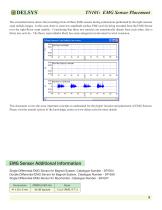
The screenshot below shows the recordings from all three EMG sensors during contractions performed by the right extensor carpi radialis longus. In this case, there is some low amplitude surface EMG activity being recorded from the EMG Sensor over the right flexor carpi radialis. Considering that these two muscles are anatomically distant from each other, this is likely true activity. The flexor carpi radialis likely has some antagonist involvement in wrist extension. This document covers the most important concepts to understand for the proper location and placement of EMG Sensors. Please visit...
Open the catalog to page 10All Delsys catalogs and technical brochures
 EMG Signal Analysis
5 Pages
 EMG Signal Quality
6 Pages
 Delsys Product Catalog (2021)
19 Pages

- Analysis software
- Tablet computer software
- Tablet PC software
- Control software
- Windows software
- Monitoring software
- Acquisition software
- Design software
- Software module
- Data management software
- Recording software
- Server-based software
- Data analysis software
- EMG system
- Module
- Analysis software module
- Medical software module
- Portable EMG
- Electrocardiography software
- Modeling software