- Catalogs
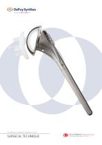
- Depuy Synthes
- GLOBAL Advantage Surgical Technique
GLOBAL Advantage Surgical Technique

GLOBAL Advantage Surgical Technique
The GLOBAL ADVANTAGE Shoulder System is engineered for global application, offering various sizes of glenoids, humeral bodies, and heads. It addresses challenges in shoulder arthroplasty such as surgical exposure, soft tissue balancing, and component fixation. The system reduces bone cement usage and enhances component stability through specialized techniques and instrumentation.
The glenoid component is designed to restore the articulating surface with minimal bone compromise. It features a larger diametral curvature than the humeral head for translation and shock absorption, with custom spherical reaming and five-peg or keel fixation for stability.
The humeral component includes a body and head, designed to fit a wide range of humeral canals with six available sizes. Made of high-strength titanium alloy, it ensures biocompatibility and optimal alignment and stability through a system of cutting and broaching instruments.
With 15 standard and 8 eccentric head components, the system ensures complete coverage of the proximal humerus. The heads are made of cobalt chrome alloy for superior wear characteristics and are joined to the body by a reverse taper lock.
The surgical technique focuses on soft tissue balancing, tendon lengthening, and capsular releases, providing flexibility to match various anatomic requirements while ensuring secure fixation, bone conservation, and mechanical optimization.
Patient Positioning: The patient is placed in a semi-Fowler position.
Surgical Incision: An incision is made from the clavicle over the coracoid down the arm's anterior aspect.
Pectoralis Major Tendon Release: The upper 25% of the tendon is released for joint exposure.
Nerve Identification: The musculocutaneous and axillary nerves are identified and protected.
Subscapularis Tendon Release: The tendon is released from the lesser tuberosity.
Capsule Release and Humeral Head Resection: The capsule is released to facilitate humeral head exposure and resection.
The document outlines a postoperative protocol to ensure optimal recovery and function of the shoulder arthroplasty.
This document details the surgical technique for humeral head removal and replacement using the DePuy Synthes GLOBAL ADVANTAGE system, including procedures for head removal, medullary canal preparation, and glenoid preparation.
Describes using an electro-cautery blade and plastic template to prevent arcing and ensure proper prosthesis seating, with strategies for posterior glenoid erosion.
An alternative guide involves creating a pilot hole and reaming the intramedullary canal for proper head removal and retroversion alignment.
Advises reaming the medullary canal using a T-handle to avoid excessive bone removal, determining the stem size for the prosthesis.
The broach size is determined by the reamer used, ensuring proper fit by aligning broach fins with osteotome tracks.
Guidelines for selecting the appropriate glenoid prosthesis size and type, emphasizing matching with the humeral head.
Provides comprehensive guidance on humeral head removal and replacement procedures, emphasizing precision in alignment and sizing for successful outcomes.
Three types of driver shafts are available: straight, articulated, or 45-degree angle. The drill bit features a sharp pilot tip to prevent wandering, with five sizes of standard reamers and a sixth for hemiarthroplasty preparation.
Glenoid Preparation: Central hole drilling using a central drill guide, avoiding over-reaming for proper peg hole alignment.
Pegged Glenoid Trial: Peripheral drill guide used for pegged glenoid prosthesis trial insertion.
Keeled Glenoid Trial: Additional steps for keel slot creation using templates and drilling.
Hand reaming is recommended initially, with power reaming as needed. Eccentric trial heads are used for off-center cavities.
Pegged Glenoid: Cement is used for prosthesis securing, avoiding excessive cement.
Keeled Glenoid: Similar cementing techniques with emphasis on a good cement mantle.
The final prosthesis is slightly larger than the trial broach for a firm press-fit, with bone graft or cement used if necessary.
Ensure proper soft tissue balance with the trial humeral head, using eccentric heads if needed. Avoid excessive cement to prevent uneven seating and potential damage.
Procedure for using cancellous bone grafts in shoulder arthroplasty, with options for cement or press-fit technique based on conditions like fractures or osteoporosis.
Cement is generally not placed deep in the canal unless defects are present, with manual prosthesis insertion using a Delrin tipped impactor.
Instructions for removing humeral head and cemented prosthesis using specific tools.
Describes reducing the humeral head into the glenoid fossa and securing the subscapularis tendon for immediate passive movement post-surgery.
Wound irrigation with antibiotic solution and closed wound drainage system recommended, with specific suture technique for cosmetic closure and cold wrap for pain reduction.
Includes removing shoulder immobilizer on the day of surgery, passive flexion exercises, and gentle daily activities, with a detailed exercise regimen and follow-up guidelines for recovery and strengthening.
Emphasizes lifelong rehabilitation for shoulder strength and flexibility, with specific exercises for deltoid and rotator cuff muscles.
Catalog excerpts
This publication is not intended for distribution in the USA. SURGICAL TECHNIQUE
Open the catalog to page 1
The GLOBAL ADVANTAGE™ Humeral Body 2 The GLOBAL ADVANTAGE Humeral Head 2 Pectoralis Major Tendon Release 5 Anterior Humeral Circumflex Vessels Management 5 Subscapularis Tendon Release 7 Capsule Release and Humeral Head Resection 8 Humeral Head Resection 8 Technique for Head Removal Using the Intramedullary Humeral Resection Guide 10 Sizing the Resected Humeral Head 12 Medullary Canal Preparation and Broaching the Humerus 13 Medullary Canal Reaming 13 Using the Body Sizing Osteotome 14 Pegged Glenoid Trial 18 Keeled Glenoid Trial 19 Humeral Head Trials...
Open the catalog to page 2
Design Rationale The multiple sizes of the glenoids, humeral bodies and heads allow the GLOBAL ADVANTAGE Shoulder System to be used worldwide. Its design is based on the detailed investigations of the structure and mechanics of normal and prosthetic glenohumeral joints conducted at the University of Texas at San Antonio, University of Washington, University of Pennsylvania and DePuy Synthes, Warsaw, Indiana. The challenges encountered by shoulder arthroplasty surgeons include surgical exposure, soft tissue balancing and component fixation. The instruments, technique and components of this arthroplasty...
Open the catalog to page 3
The Technique Recognising that a successful shoulder arthroplasty is critically dependent on soft tissue balancing, this document provides a detailed guide to the techniques of tendon lengthening and capsular releases, that are integral parts of this procedure. These steps cannot be effected with jigs and guides, but rather require an understanding of the principles of shoulder mechanics. With the aim that each shoulder arthroplasty is adapted to the patient’s combination of soft tissue and bone anatomy, the system is designed to maximise the surgeon’s flexibility in matching a wide variety of...
Open the catalog to page 4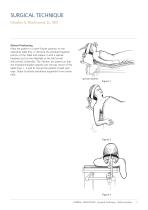
SURGICAL TECHNIQUE Charles A. Rockwood, Jr., MD Patient Positioning Place the patient in a semi-Fowler position on the operating table (Fig. 1). Remove the standard headrest portion of the table and replace it with a special headrest such as the Mayfield or the McConnell (McConnell, Greenville, TX). Position the patient so that the involved shoulder extends over the top corner of the table (Figs. 1, 2 and 3). Secure the patient’s head with tape. Drape to isolate anesthesia equipment from sterile field. Special headrest Figure 3 GLOBAL ADVANTAGE Surgical Technique DePuy Synth
Open the catalog to page 5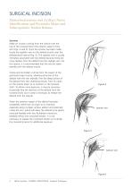
SURGICAL INCISION Musculocutaneous and Axillary Nerve Identification and Pectoralis Major and Subscapularis Tendon Release Incision Make an incision running from the clavicle over the top of the coracoid down the anterior aspect of the arm (Figs. 4 and 5). Once the incision has been made, locate the cephalic vein on the deltoid muscle near the deltopectoral interval (Fig. 6). The cephalic vein is usually intimately associated with the deltoid because there are many feeders from the deltoid into the cephalic vein. For this reason, it is recommended that the vein be taken laterally with the deltoid...
Open the catalog to page 6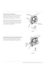
Pectoralis Major Tendon Release Release the upper 25 percent of the pectoralis major tendon from its insertion on the humerus with an electro-cautery cutting blade. This will aid in the exposure of the inferior aspect of the joint (Fig. 7). Richardson retractor If the patient has marked internal rotation contracture, release most of the pectoralis major tendon from its insertion. This tendon release should not be repaired at the completion of the operation since it will limit external rotation postoperatively. Pectoralis major tendon Anterior Humeral Circumflex Vessels Management Isolate, clamp...
Open the catalog to page 7
NERVE IDENTIFICATION Musculocutaneous Nerve It is important to identify the musculocutaneous and axillary nerves. Palpate the musculocutaneous nerve as it comes from the plexus into the medial and posterior aspect of the conjoined tendon (Fig. 9). Usually, the nerve penetrates the muscle approximately 4 to 5 cm - or - 3.8 to 5.1 cm. down from the tip of the coracoid, but in some instances the nerve has a higher penetration into the conjoined muscle tendon unit. Remember the proximity of this nerve to the tendon during the retraction of the conjoined tendon. Coracoid process Conjoined tendon Musculocutaneous...
Open the catalog to page 8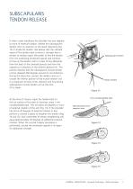
SUBSCAPULARIS TENDON RELEASE If when under anesthesia the shoulder has zero degrees or more of external rotation, release the subscapularis tendon from its insertion on the lesser tuberosity (Fig. 12) or divide the tendon. We believe that the ultimate repair of the tendon back to bone is stronger than a tendon to tendon repair. We prefer to free the tendon from the underlying thickened capsule and continue to free up the tendon until it is clear of any adhesions from the back of the coracoid process and from the capsule as it attaches on the anterior glenoid rim. This process requires that the...
Open the catalog to page 9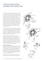
CAPSULE RELEASE AND HUMERAL HEAD RESECTION Occasionally, the capsule will be released from the neck of the humerus with the subscapularis tendon. If that occurs, dissect the anterior capsule from the posterior surface of the subscapularis to maintain a free, dynamic subscapularis tendon. Use a Scoffield retractor to retract the previously identified axillary nerve anteriorly/ inferiorly away from the inferior capsule. Externally rotate the arm, which will place tension on the capsule, and then release the capsule from its attachment to the humerus all the way down inferiorly to at least the six...
Open the catalog to page 10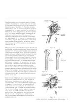
Curved crego retractor Place the template along the anterior aspect of the arm parallel to the shaft of the humerus, and mark the angle at which the head will be removed with an osteotome or the electro-cautery blade (Fig. 21). The plastic template prevents arcing from the electro-cautery knife. Use of the template ensures the proper seating of the prosthesis on the bone (Fig. 22). In many instances, the inferior portion of the mark will be above the inferior osteophyte of the flattened and deformed head of the humerus. If the resection is made in line with an articular surface which is in varus,...
Open the catalog to page 11All Depuy Synthes catalogs and technical brochures
 Titanium Sternal Fixation System
34 Pages
 Small Battery Drive II
4 Pages
 Introducing The Variable Angle
12 Pages

Archived catalogs
 MatrixRIB®FixationSystem
86 Pages
 HEALIX ADVANCE
4 Pages
 HEALIX Anchor™ 3.4 mm
2 Pages
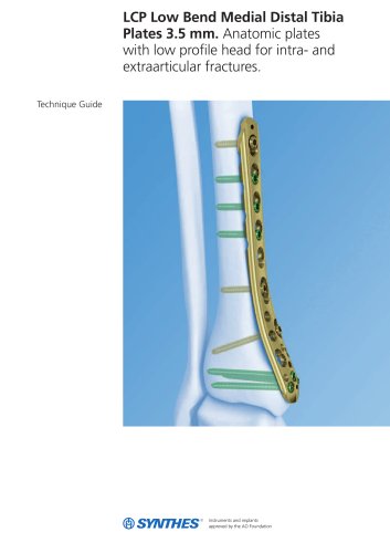
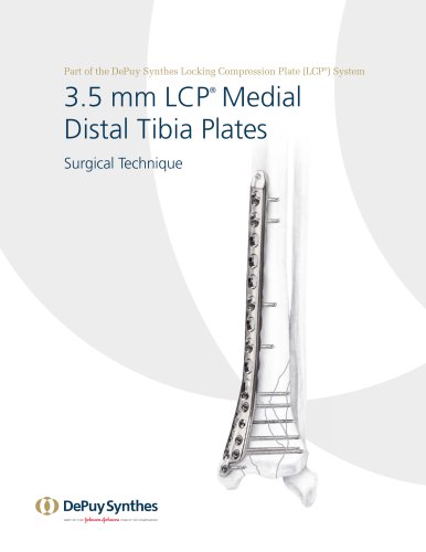 3.5 mm LCP™ Medial
15 Pages
 RADIUS OF CURVATURE
3 Pages
 Building on Success
16 Pages
 2.0 mm LCP® Distal Ulna Plate
20 Pages
 2.4 mm VA LCP™
4 Pages
 Mandible Trauma Solutions
2 Pages
 Power line II
4 Pages
 Concorde
28 Pages
 LCP Intercarpal
31 Pages
 LCS® COMPLETE™
2 Pages
 Synthes TPLO.
8 Pages
 SynFix-LR System
56 Pages
 ATB Anterior Tension Band Plate
32 Pages
 CONDUIT™
15 Pages
 Brochure_FINAL
2 Pages
 DePuy Synthes
81 Pages
 Anspach
3 Pages
 Orthopedic Foot Instruments
32 Pages
 PINNACLE® Hip Solutions
12 Pages
 Corail
24 Pages
 S-ROM® NOILES™
68 Pages
 TRI-LOCK® Product Rationale
12 Pages
 Reclaim Surgical Technique
44 Pages
 Speed
Speed2 Pages
 attune
80 Pages
 HAMMERLOCK® 2
2 Pages
 DePuy Glenoid Solutions
2 Pages
 Trauma Solutions. Elbow
4 Pages
 Polar
Polar4 Pages
 Alveolar Distractor.
4 Pages
 Piezoelectric System
4 Pages
 Air Power Line II
6 Pages
 LCP Clavicle Hook Plate
4 Pages
 TruMatch Pin Guides
16 Pages
 P F N A
8 Pages
 SKILL, DEDICATION,
16 Pages
 Orthopaedics. Overview
20 Pages
 DURALOC
16 Pages
 Marathon Cemented Cup
20 Pages
 REEF Surgical Technique
16 Pages
 MatrixNEURO
8 Pages
 Anspach XMax
4 Pages
 Anspach eMax 2 Plus
4 Pages
 Small Electric Drive
4 Pages
 Air Pen Drive
4 Pages
 Colibri II
4 Pages
 Spine
Spine25 Pages
 Expert Hindfoot Arthrodesis Nail
48 Pages
 LCP Distal Fibula Plates
32 Pages
 TomoFix
60 Pages
 Expert Tibial Nail PROtect
16 Pages
 Expert Tibia Nail
84 Pages
 Sacral Bars
16 Pages
 Pelvic C-Clamp
20 Pages
 Low Profile Pelvic System
16 Pages
 Proximal Femoral (Hook) Plate
24 Pages
 LCP
LCP24 Pages
 PFNA
PFNA112 Pages
 HCS 1.5, 2.4, 3.0
36 Pages
 LCP Wrist Fusion
32 Pages
 LCP Compact Hand
28 Pages
 VA-LCP Elbow
48 Pages
 Distal Radius
44 Pages
 Olecranon
30 Pages
 LCP Hook Plate
28 Pages
 DHP & Olecranon
4 Pages
 LCP S-A
4 Pages
 Epoca
Epoca4 Pages
 Philos
32 Pages
 MultiLoc
68 Pages


- DePuy Synthes bone plate
- Compression plate
- Metallic compression plate
- Locking compression plate
- Titanium compression plate
- Distal compression plate
- Orthopedic surgery instrument kit
- Interbody fusion cage
- Sterilization container
- Instrument sterilization container
- Arthrodesis nail
- Bone substitute
- Metallic intramedullary nail
- Anterior interbody fusion cage
- Femoral stem
- Arthrodesis plate
- Orthopedic surgery bone substitute
- Femoral intramedullary nail
- Suture anchor
- Knee prosthesis