
- Catalogs
- Depuy Synthes
- TomoFix


TomoFix

TomoFix
The TomoFix Medial High Tibial Plate (MHT) is designed for medial high tibial osteotomies, providing stability and support for joint-preserving knee surgeries. It features a locking compression plate system that ensures angular stability and rapid osseous healing.
Indications include open-wedge and closed-wedge osteotomies of the medial proximal tibia, unicompartmental gonarthrosis with malalignment, and varus or valgus deformity. It is contraindicated in cases of inflammatory arthritis.
The TomoFix system is suitable for both open and closed-wedge osteotomies. The guide focuses on open-wedge osteotomy procedures, emphasizing the importance of preoperative planning using the Miniaci method.
Preoperative planning involves determining the mechanical axis and hinge point on weight-bearing x-rays. The surgical approach includes positioning the patient supine, marking anatomical landmarks, and making a skin incision. Care is taken to avoid damage to the saphenous nerve.
The procedure involves placing Kirschner wires to guide the osteotomy, performing a biplanar osteotomy with an oscillating saw, and ensuring proper orientation and depth of cuts. The ascending osteotomy cut is crucial for maintaining tibial slope inclination.
The TomoFix plate is positioned using guiding blocks and spacers to ensure correct alignment and compression on the lateral hinge. The plate's design allows for easy insertion and stable fixation.
Postoperative care involves monitoring healing and planning for eventual implant removal once the osteotomy has healed.
The guide includes detailed specifications of plates, screws, and instruments used in the procedure, emphasizing the importance of using the correct components for successful outcomes.
The TomoFix MHT system provides a reliable solution for medial high tibial osteotomies, offering stability and support for knee joint preservation. Proper training and adherence to the technique guide are essential for optimal results.
Instruments include various widths of TomoFix Osteotomy Chisels, a TomoFix Bone Spreader, Bone Spreader forceps, and alignment tools such as the Alignment Rod and TomoFix Osteotomy Gap Measuring Device. Additional tools include drill bits, drill guides, torque limiters, and screwdrivers for precise fixation.
- Osteotomy Opening: Use chisels to open the osteotomy, ensuring the insertion depth matches the cutting depth. Maintain guide wires in place to prevent fracturing.
- Spreading the Osteotomy: Gradually spread the osteotomy to avoid fractures, using either chisels, a bone spreader, or forceps. Ensure symmetrical opening and sufficient release of the medial collateral ligament.
- Checking Correction: Constantly monitor leg alignment and osteotomy height using the alignment rod and image intensifier. Adjust as necessary to match the preoperative plan.
- Plate Insertion and Fixation: Insert the plate subcutaneously, ensuring proper alignment. Secure with Kirschner wires and self-tapping locking screws, using a torque limiter for final tightening.
- Lag Screw Insertion: Insert a lag screw to compress the lateral hinge, ensuring no correction loss and avoiding soft tissue compression.
- Distal Plate Fixation: Use a monocortical self-drilling locking screw for distal fixation, replacing spacers with locking head screws as needed.
- Ensure the osteotomy is spread slowly to prevent fractures.
- Monitor the osteotomy with an image intensifier in two planes to avoid malrotation and destabilization.
- Use a torque limiter to prevent cold welding of screws and ensure proper tightening.
- Always perform checks with the leg in full extension to ensure accurate alignment.
- Use the alignment rod for checking purposes, but confirm with a full leg x-ray for absolute accuracy.
Early functional postoperative treatment with partial load bearing is advised, along with physiotherapy and preventive measures against thrombosis. Implant removal is generally not necessary unless desired after complete healing.
A comprehensive list of instruments required for the procedure is provided, including drill bits, torque limiters, screwdrivers, and optional instruments like osteotomy chisels and bone spreaders.
Synthes offers chronOS wedges, a synthetic osteoconductive material that can be used in conjunction with bone marrow aspirate to enhance bone healing. The material is resorbable and can be trimmed to fit, providing optimal conditions for faster bone healing.
- Enhances osteogenic potential using autologous bone marrow.
- Facilitates efficient intraoperative impregnation of chronOS products with the patient's bone marrow or blood.
- Designed for ease of use and minimal invasiveness.
- Compatible with the chronOS Perfusion System for direct bone marrow transfer into perfusion devices containing chronOS implants.
- chronOS Wedges available in semicircular shapes with angles of 7°, 10°, and 13° and dimensions of 25x35 mm.
- Wedges also available with Perfusion System compatibility.
- Studies on primary stability and effects of high tibial osteotomy on knee cartilage pressure.
- Research on osteopromotion using _-TCP/Bone Marrow Hybrid Implants in spine surgery.
- Various studies and bibliographies on osteotomies around the knee, surgical techniques, and fixation devices.
- Technique guides are available as PDF files on the Synthes website.
- All products and techniques are subject to modification and are trademarks of Synthes, Inc. or its affiliates.
Catalog excerpts
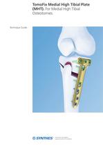
TomoFix Medial High Tibial Plate (MHT). For Medial High Tibial Technique Guide
Open the catalog to page 1
Image intensier control Warning This description alone does not provide sufficient background for direct use of the product. Instruction by a surgeon experienced in handling this product is highly recommended. Reprocessing, Care and Maintenance of Synthes Instruments For general guidelines, function control and dismantling of multi-part instruments, please refer to: www.synthes.com/reprocessing
Open the catalog to page 2
ntroduction TomoFix Medial High Tibial Plate (MHT) 2 Surgical Technique General Remarks 5 Positioning and Fixation of the Plate 23 Postoperative Treatment and Implant Removal 43 Product Information Plates 44 Also Available from Synthes: chronOS Wedges 53 TomoFix Medial High Tibial Plate (MHT) Technique Guide Synthes 1
Open the catalog to page 3
TomoFix Medial High Tibial Plate (MHT). For Medial High Tibial Osteotomies. Features and Benefits Higher stability Four locking screws in the plate head Long shaft portion Evenly transmits the occurring forces into the tibial shaft Compression of the lateral hinge A lag screw pulls the distal osteotomy segment towards the plate… 2 Synthes …and forces the plate into suspension, creating an elastic preload… TomoFix Medial High Tibial Plate (MHT) Technique Guide …which imposes pressure upon the lateral hinge.
Open the catalog to page 4
Easy plate insertion Compression of the lateral hinge TomoFix technique allows pretension- ng of the plate using cortex screw and TomoFix Knee Osteotomy System TomoFix Tibial Head Plate medial, proximal wedge high tibial - Increased plate strength preload technique stable bridging small stature versions TomoFix Tibial Head Plate lateral, proximal wedge osteotomies stable fixation - Available in right and left TomoFix Femoral Plate medial, distal stable fixation - Available in right and left TomoFix Femoral Plate lateral, distal wedge osteotomies stable fixation - Available in right and left TomoFix...
Open the catalog to page 5
Indications and Contraindications Indications: − Open-wedge and closed-wedge osteotomies of the medial proximal tibia − Unicompartmental medial or lateral gonarthrosis with malalignment of the proximal tibia − Idiopathic or posttraumatic varus or valgus deformity of the proximal tibia Contraindication: − Inflammatory arthritis 4 Synthes TomoFix Medial High Tibial Plate (MHT) Technique Guide
Open the catalog to page 6
General Remarks Open-wedge corrective tibia osteotomies are increasingly being used for joint-preserving surgery of the knee joint. The TomoFix knee osteotomy system is based on the Locking Compression Plate system (LCP) and enables angular-stable connections between the screw and plate. This angular stability allows the stable fixation of an open-wedge osteotomy and hence its rapid osseous healing. Note: Plan the type and position of the osteotomy. The TomoFix medial high tibial plate is suitable for both open and closed-wedge osteotomies. This technique guide will explain the procedure of an...
Open the catalog to page 7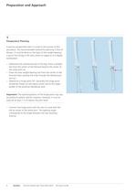
Preparation and Approach 1 Preoperative Planning A precise preoperative plan is crucial to the success of this procedure. The recommended method for planning is that of Miniaci. It must be done on the basis of the weight-bearing x-ray of the full leg in AP view, either on paper or at a digital workstation. − Determine the mechanical axis of the leg: Draw a straight line from the center of the femoral head to the center of the ankle joint (a). − Draw the new weight-bearing line from the center of the femoral head, passing the knee through the desired position (a’). − Determine a hinge point (H)....
Open the catalog to page 8
- Determine the entry point of the transverse osteotomy. It ies just above the pes anserinus. Make sure there is still enough space for the plate head, so that the screw in hole D can be inserted without protruding into the wedge. De- pending on the determined opening angle and the length of the osteotomy cut (mediolateral diameter of the os- teotomy) the corresponding opening height can be de- rived from Hernigou's trigonometric chart. Trigonometric chart Correction angle Note: These instructions alone do not replace in-depth train- ng in planning for osteotomies. It only serves as a general...
Open the catalog to page 9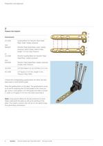
312.924 Guiding Block for TomoFix Tibial Head Plate, small, medial, proximal 440.831 TomoFix Tibial Head Plate, small, medial, proximal, shaft 4 holes, head 4 holes, length 112 mm. Pure Titanium 312.926 TomoFix Guiding Block for TomoFix Tibial Head Plate, medial, proximal 440.834 TomoFix Tibial Head Plate, medial, proximal, 4 holes. Pure Titanium 323.042 LCP Drill Sleeve 5.0, for Drill Bits 0 4.3 mm Choose the corresponding guiding block for either the stan- dard sized or small TomoFix plate. Place the guiding block on the plate. The guiding block serves as an aid for attaching the LCP drill...
Open the catalog to page 10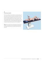
3 Positioning of patient Perform the surgery with the patient in a supine position. Attach a lateral support and foot pad to the operating table so that the leg can be easily positioned in 90° flexion and in full extension. Position the patient so that the hip, knee and ankle joint can be visualized with the image intensifier. Lower the contralateral leg at the hip joint to facilitate access to the medial proximal tibia. The sterile draping also exposes the iliac crest so that the leg axis can be checked intraoperatively. A sterile tourniquet can be used, but is not mandatory. Note: Allow enough...
Open the catalog to page 11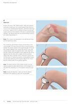
Preparation and Approach 4 1 Approach Position the knee in 90° flexed position. Mark the anatomic landmarks (medial joint line, cranial border of pes anserinus, course of the medial collateral ligament, and tibial tuberosity) on the skin. Make a 6 – 8 cm long skin incision running from a point anterior to the insertion of the pes in a posterocranial direction. The incision ends over the posteromedial corner of the medial tibial plateau.(1) Note: This incision runs essentially in line with the skin lines and the saphenous nerve. First, divide the subcutaneous tissues and the fascia at the cranial...
Open the catalog to page 12All Depuy Synthes catalogs and technical brochures
 Titanium Sternal Fixation System
34 Pages
 Small Battery Drive II
4 Pages
 Introducing The Variable Angle
12 Pages

Archived catalogs
 MatrixRIB®FixationSystem
86 Pages
 HEALIX ADVANCE
4 Pages
 HEALIX Anchor™ 3.4 mm
2 Pages
 3.5 mm LCP™ Medial
15 Pages
 RADIUS OF CURVATURE
3 Pages
 Building on Success
16 Pages
 2.0 mm LCP® Distal Ulna Plate
20 Pages
 2.4 mm VA LCP™
4 Pages
 Mandible Trauma Solutions
2 Pages
 Power line II
4 Pages
 Concorde
28 Pages
 LCP Intercarpal
31 Pages
 LCS® COMPLETE™
2 Pages
 Synthes TPLO.
8 Pages
 SynFix-LR System
56 Pages
 ATB Anterior Tension Band Plate
32 Pages
 CONDUIT™
15 Pages
 Brochure_FINAL
2 Pages
 DePuy Synthes
81 Pages
 Anspach
3 Pages
 Orthopedic Foot Instruments
32 Pages
 PINNACLE® Hip Solutions
12 Pages
 Corail
24 Pages
 S-ROM® NOILES™
68 Pages
 TRI-LOCK® Product Rationale
12 Pages
 Reclaim Surgical Technique
44 Pages
 Speed
Speed2 Pages
 attune
80 Pages
 HAMMERLOCK® 2
2 Pages
 DePuy Glenoid Solutions
2 Pages
 Trauma Solutions. Elbow
4 Pages
 Polar
Polar4 Pages
 Alveolar Distractor.
4 Pages
 Piezoelectric System
4 Pages
 Air Power Line II
6 Pages
 LCP Clavicle Hook Plate
4 Pages
 TruMatch Pin Guides
16 Pages
 P F N A
8 Pages
 SKILL, DEDICATION,
16 Pages
 Orthopaedics. Overview
20 Pages
 DURALOC
16 Pages
 Marathon Cemented Cup
20 Pages
 REEF Surgical Technique
16 Pages
 MatrixNEURO
8 Pages
 Anspach XMax
4 Pages
 Anspach eMax 2 Plus
4 Pages
 Small Electric Drive
4 Pages
 Air Pen Drive
4 Pages
 Colibri II
4 Pages
 Spine
Spine25 Pages
 Expert Hindfoot Arthrodesis Nail
48 Pages
 LCP Distal Fibula Plates
32 Pages
 Expert Tibial Nail PROtect
16 Pages
 Expert Tibia Nail
84 Pages
 Sacral Bars
16 Pages
 Pelvic C-Clamp
20 Pages
 Low Profile Pelvic System
16 Pages
 Proximal Femoral (Hook) Plate
24 Pages
 LCP
LCP24 Pages
 PFNA
PFNA112 Pages
 HCS 1.5, 2.4, 3.0
36 Pages
 LCP Wrist Fusion
32 Pages
 LCP Compact Hand
28 Pages
 VA-LCP Elbow
48 Pages
 Distal Radius
44 Pages
 Olecranon
30 Pages
 LCP Hook Plate
28 Pages
 DHP & Olecranon
4 Pages
 LCP S-A
4 Pages
 Epoca
Epoca4 Pages
 Philos
32 Pages
 MultiLoc
68 Pages




- DePuy Synthes bone plate
- Metallic compression plate
- Titanium compression plate
- Orthopedic surgery instrument kit
- Distal compression plate
- Interbody fusion cage
- Sterilization container
- Instrument sterilization container
- Arthrodesis nail
- Bone substitute
- Metallic intramedullary nail
- Femoral stem
- Anterior interbody fusion cage
- Arthrodesis plate
- Orthopedic surgery bone substitute
- Femoral intramedullary nail
- Knee prosthesis
- Metallic arthrodesis plate