- Products
- Catalogs
- News & Trends
- Exhibitions
OP 3D
1 /9Pages

OP 3D
1 /9Pages
Catalog excerpts

Imaging innovations in one device
Open the catalog to page 1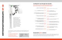
Leading the way through the decades For more than 60 years, the name of ORTHOPANTOMOGRAPH system has stood for ultimate reliability and clinically correct maxillofacial imaging. Professor Y.V. Paatero publishes his first paper on Panoramic Tomography. The first dental panoramic x-ray, ORTHOPANTOMOGRAPH OP1, is developed. • Implantology • Endodontics • Impactions • TMJ Volumetric Tomography (VT) is developed to maximize the performance of an ORTHOPANTOMOGRAPH unit. CBCT era begins. ORTHOPANTOMOGRAPH OP300, the most comprehensive 3-in-1 platform, is launched to celebrate 50 years of ORTHOPANTOMOGRAPH...
Open the catalog to page 2
New level of control and flexibility With the OP 3D system the FOV location can be controlled easily and accurately. SMARTVIEW™ 2.0 userinterface offers two-dimensional scout images prior to the actual CBCT examination. Users can verify the exact FOV location with the ability to adjust automatically based on the selection. This limits the need for retakes and can help lower dosage and follow ALARA (As Low As Reasonably Achievable) radiation protocols. Every feature of the OP 3D system is designed to increase practice efficiency. Preparing the unit for a scan is fast with an intuitive patient...
Open the catalog to page 3
Tools for professionals One size does not have to fit all. The OP 3D system offers efficient tools for optimizing the patient dose with its ability to allow the clinician to select the best resolution, FOV size, and region of interest. Clearer images with MAR technology FOV 6 x 9 cm Covers the complete lower or upper jaw with opposing occlusion. To provide the highest level of image quality, the Metal Artifact Reduction (MAR) is readily activated with all the FOV sizes of OP 3D. MAR is optimized to aid in all cases ranging from endodontics and implants to maxillofacial imaging. Noise reduction...
Open the catalog to page 4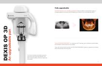
Fully upgradeable The OP 3D panoramic unit is completely upgradeable. Choose the addition of cephalometric imaging, or completely upgrade and choose to add 3D imaging to your practice for even more diagnostic options. The first ORTHOPANTOMOGRAPH™ unit, introduced over 50 years ago, was a revolutionary ground breaker and pacesetter for dental panoramic X-ray imaging. Today, with more than 60,000 units sold, the ORTHOPANTOMOGRAPH systems are regarded as the leading name and benchmark in the X-ray world. The 9-second standard and pediatric panoramic scan times provide clear image definition, with...
Open the catalog to page 5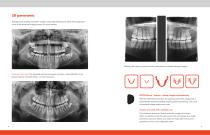
2D panoramic Standard and pediatric panoramic images, along with bitewing and lateral TMJ projections cover all the extraoral imaging needs of a busy practice. Bitewing-like view is a quick and easy alternative to intraoral bitewing imaging. 9-Second scan time: The standard panoramic program provides a clear definition of the dental anatomy, including TMJs—in only 9 seconds. ORTHOfocus™ feature—sharp images automatically With the ORTHOfocus feature, the optimum panoramic image layer is automatically obtained enabling forgiving patient positioning. The result is consistent image quality every...
Open the catalog to page 6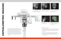
Lateral cephalometric images provide Pediatric lateral images with reduced PA cephalometric images offer great rich anatomical details with exceptional height allow one to minimize the dose. details — thanks to the powerful visibility of the soft tissue borderline. dedicated X-ray source. Carpus imaging-information to determine Lateral cephalometric programs for adult and pediatric patients with adjustable 16 to 26 cm fields width. The innovative, patented ORTHOceph™ Plus design of the OP 3D system takes cephalometric imaging workflow to a new level. The OP 3D system provides needed protocols...
Open the catalog to page 7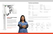
Technical specifications 2D / Cephalometric 2D / Panoramic Image receptor Image receptor Pixel size (sensor & image) 99 μm Pixel size (sensor & image) 99 μm Tube voltage Tube voltage Tube current Tube current Scan time Scan time Image field height Image field height Imaging programs Standard, Segmented, Pediatric, Lat TMJ, Image field width Imaging programs Lateral and Pediatric Lateral with an adjustable field width, Posterior-Anterior (PA), Carpus*. 3D / CBCT Image detector Tube focal spot Image voxel size DICOM** support Available as a software option. Tube voltage Tube current Scan time Image...
Open the catalog to page 8
About DEXIS DEXIS innovation is nothing new. After all, our recognized, trusted products are built on over 200 years of dental imaging expertise, combining leading brands including i-CAT™, Gendex™, Instrumentarium, SOREDEX™, and NOMAD™ Pro 2. Today, over 150,000 offices trust DEXIS products around the world. DEXIS now includes a full portfolio of products including CBCT and intraoral scanners, our legacy digital sensors and handheld x-ray system and DTX Studio™ Clinic, the next generation software. This complete digital solution works seamlessly together as well as with other systems to enhance...
Open the catalog to page 9All DEXIS catalogs and technical brochures
 OP 2D
OP 2D8 Pages
 IXS
IXS4 Pages
 Nomad Pro 2
8 Pages
 DEXISTM Titanium by KaVo
12 Pages
- Visualization software
- Radiology software
- Tablet computer software
- Tablet PC software
- Flat panel detector
- Control software
- Windows software
- Diagnostic software
- Planning software
- Cloud-based software
- Dental software
- Design software
- Dental radiography system
- Acquisition software
- Capture software
- Software module
- AI-assisted software
- Artificial intelligence software
- Digital dental radiography system
- 3D scanner