- Catalogs
- DIAL Medicali
- T-FixUp

- Products
- Catalogs
- News & Trends
- Exhibitions
T-FixUp
T-FixUp
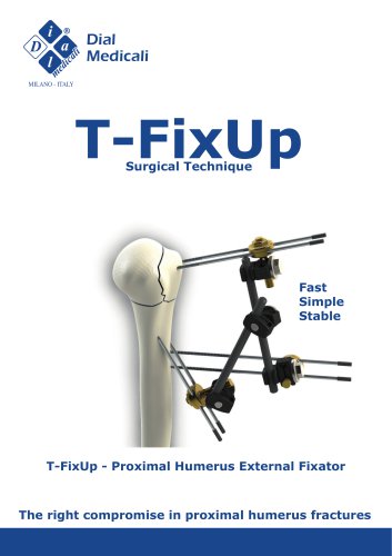
The T-FixUp is an external fixator system designed for stabilizing proximal humerus fractures. It consists of various components assembled on a PinFix pin inserted into the bone, following a specific surgical technique.
Key Components
- PinFix 2.5x300mm thread 60mm
- Pin clamp
- Bar to bar 6mm clamp
- Carbon bars of various lengths
- 10mm wrench
Preoperative Planning
Essential radiographic examinations include anteroposterior, axial, Y, Grashey, and transthoracic projections. A CT scan with multiplanar and 3D reconstruction is recommended for complex fractures.
Patient Positioning
The patient is initially positioned supine for reduction maneuvers, then in a beach chair position for surgery. Proper X-ray projections should be ensured before surgery.
Operating Field Preparation
Shoulder washing with chlorhexidine and proper draping to expose key anatomical landmarks is recommended. The arm should be free to move.
Anatomical Landmarks
Identifying the coracoid process and acromial edge is crucial. A longitudinal line is drawn for pin insertion, considering the axillary nerve's path.
Closed Reduction Techniques
Reduction maneuvers aim to realign bone segments. In difficult cases, a K-wire can be used as a joystick for better reduction.
Percutaneous Fixation
- Make an incision and insert the first pin with fluoroscopic guidance, considering anatomical parameters.
- Use a pin clamp for parallel insertion of the second pin.
- Insert the third pin from the greater tuberosity apex.
- Insert the fifth and sixth pins in the shaft.
- Connect pin clamps with carbon bars to form a stable construct.
- Check construct stability intra-operatively.
Open Reduction
In cases of severe comminution, open reduction may be necessary using deltoid-pectoral or MacKenzie approaches.
Device Removal
The fixator is typically removed after 40-45 days using a power drill.
Additional Applications
The T-FixUp can be used in pediatric cases, for radius or ulna fixation, and in complex circular fixation systems.
Components List
A detailed list of components, including PinFix pins, clamps, bars, and wrenches, is provided.
Acknowledgments
Thanks to Prof. F. D'Angelo, Dr. A. Pautasso, and Dr. M. Puricelli for their contributions.
Catalog excerpts
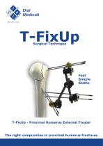
Dial Medicali MILANO - ITALY T-FixUp Surgical Technique Fast Simple Stable T-FixUp - Proximal Humerus External Fixator The right compromise in proximal humerus fractures
Open the catalog to page 1
The T-FixUp fixator is made up of various components which, assembled together on PinFix pin, inserted into the bone, according to the surgical technique reported below, forms an external osteosynthesis system capable of stabilizing the fracture until healing. The technique illustrates the individual procedures to obtain correct percutaneous stabilization of proximal humerus fractures. • PinFix 2.5x300mm thread 60mm (code 16-0050/NS) • Pin clamp (code 02-81090 paten pending) Note: Complete in-depth reading is recommended before using the product Radiographic examinations (anteroposterior projection,...
Open the catalog to page 2
ANATOMICAL LANDMARKS Drawing the precise points of the shoulder bony processes helps the surgeon during the percutaneous fixation technique, moreover, it reduces the exposure to the x-rays. It is important identify the coracoid process and the entire acromial edge process. Draw a longitudinal line starting from the anterolateral corner of the acromial process along the anterolateral edge of the humeral shaft (MacKenzie approach). The axillary nerve runs approximately 5-7cm transversely, it originates posteriorly and passes anteriorly into the deltoid muscle. Finally, on the same line mark the...
Open the catalog to page 3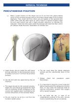
SURGICAL TECHNIQUE 1) Make a small incision on the drawn line at 10 cm from the antero-lateral corner of the acromial process and on the antero-lateral edge of the humeral diaphysis. Thus, insert the first 2.5mm pin pre-loaded on the powered drill motor while with the other hand (or with a help of a second surgeon) keep the arm push behind and with a slight shoulder abduction to maintain the reduction. Check the entry point under fluoroscope and insert the first 2.5mm pin oblique inside the bone. Parameters to consider are: a. Insert slowly the pin inside the soft tissue and pay attention to...
Open the catalog to page 4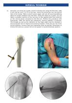
Using the pin clamp as a guide, insert it onto the wire using the first hole, slide it over the pin up to 1/2 cm to the skin. Insert the second pin into the second hole of the clamp. Make a small incision again and use all recommendations above for proper insertion into the bone. Using the clamp as a guide, you will obtain a parallel insertion of the two pins on the sagittal plane but posterior on the coronal plane compared to the first pin. Monitor the procedure with fluoroscopy. After the second pin placement, primary stability is achieved. The threads as well as the reduction of the head shaft...
Open the catalog to page 5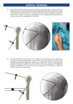
Insert the third pin starting from the greater tuberosity apex to the third lower humeral head. Use the same technique reported about the second wire: place the clamps on the third pin at 1 cm from the skin and use its slide to insert the fourth one. This pin must be more parallel on the sagittal plane but posterior on the coronal one compared to the third. Insert the fifth and sixth pins in the shaft on the drawing line at about 8-9 cm high from the anterolateral corner of the acromial process. Use the same technique reported about the second pin: place the sixth pin through the clamps inserted...
Open the catalog to page 6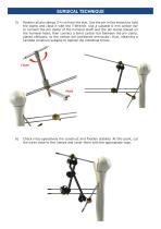
Position all pin clamps 3-4 cm from the skin. Use the pin in the wrench to hold the clamp and close it with the T-Wrench. Use a suitable 6 mm carbon bar to connect the pin clamp of the humeral shaft and the pin clamp placed on the humeral head; then connect a 6mm carbon bar between the pin clamp, placed obliquely, to the carbon bar positioned previously; thus, obtaining a Lambda construct suitable to oppose the rotational forces. Check intra-operatively the construct and fixation stability. At this point, cut the wires close to the clamps and cover them with the appropriate caps.
Open the catalog to page 7
An open reduction of the proximal humeral fracture can be performed in case of important bone comminution or difficulty reduction with external manoeuvres. Two approaches are described in literature: the classical deltoid-pectoral access and the antero-lateral trans-deltoid way (MacKenzie approach). Both approaches allow to manage the humeral fragments, but each one has advantages and disadvantages. The deltoid-pectoral way is easier to extend distally the incision and probably most of the surgeons have more confidence, because of the well-known and famous shoulder approach. However, it could...
Open the catalog to page 8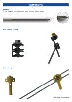
COMPONENTS PinFix 2.5 x 300mm, thread 60mm, with tip and thread detail
Open the catalog to page 9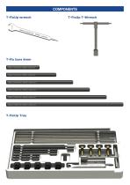
COMPONENTS T-FixUp wrench T-FixUp Tray T-FixUp T-Wrench
Open the catalog to page 10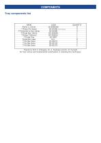
T-FixUp Pin clamp 02-81090 patent pending 4 Thanks to Prof. F. D'Angelo, Dr. A. Pautasso and Dr. M. Puricelli for their active and fundamental contribution in realizing this technique.
Open the catalog to page 11
Dial Medicali srl Viale Lombardia 6 - 20092 Cinisello Balsamo - Italy Tel. +39.02.89828078 - Fax +39.02.66104021
Open the catalog to page 12