
- Catalogs
- Dürr Dental
- VistaVox S and VistaVox S Ceph 3D from Dürr Dental

VistaVox S and VistaVox S Ceph 3D from Dürr Dental
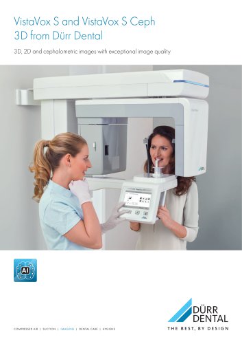
VistaVox S and VistaVox S Ceph 3D from Dürr Dental
- 3D imaging volumes are customized for the jaw arch (Ø 130 x 85 mm) and specific regions (Ø 50 x 50 mm).
- Equipped with a high-resolution CsI sensor with a pixel size of 49.5 μm for enhanced image quality.
- Radiation dose is minimized through anatomically adapted volumes and SQ mode, reducing the dose by 62% compared to HQ mode.
- Addresses tooth development issues such as hyperplasia or dysplasia.
- Useful for fractures, implant technology planning, and endodontics.
- Assists in the localization of foreign bodies and salivary stones.
- Detects pathological changes in the maxillary sinus and jawbone.
- Incorporates S-Pan technology for precise OPG image acquisition.
- Offers 17 panoramic X-ray programs, including child-specific modes and orthogonal imaging.
- Provides cephalometric exposures with six modes, including Head Lateral and Waters View.
- VistaSoft software provides intuitive handling with AI-supported features for improved diagnostics.
- Features automatic image rotation and AI-assisted nerve canal marking to enhance workflow efficiency.
- Compatible with all current Dürr Dental imaging systems and offers network capabilities for easy data access.
- Includes Comfort Bite and Universal Chin Support for stable and comfortable patient positioning.
- Facilitates simple patient positioning with alignment lights and a user-friendly touch display.
- Metal artefact reduction (MAR) for clearer 3D imaging.
- Supports digitalization of plaster casts and drill templates for 3D printing and planning.
- Offers reliable and efficient implant planning with a guided workflow.
- Facilitates simple matching of optical scan data and 3D data.
- Enables design and planning of drill templates with open source STL file export.
- Integrates with SICAT for improved digital implant workflow.
- X-ray HV Generator: Voltage range of 50–99 kV, current of 4–16 mA, and rated power of 1.6 kW.
- Image Detector: Csl CMOS photodiode array with a pixel size of 49.5 μm and an active sensor surface area of 135.8 x 36.4 mm.
- Scan Times: Ranging from 2 to 18 seconds, with specific programs for children and adolescents.
- 3D Volume Options: Various dimensions available, including Ø 130 x 50 mm and Ø 130 x 120 mm (stitched).
- Unit Dimensions: Height adjustable from 1406–2206 mm, weight of 180 kg, and installation options for wall mounting.
- Electrical Connections: Mains voltage of 200–240 V AC, frequency of 50/60 Hz, and rated power of 170 W, 2.2 kVA max.
Catalog excerpts
VistaVox S and VistaVox S Ceph 3D from Durr Dental 3D, 2D and cephalometric images with exceptional image quality DURRDENTAL COMPRESSED AIR | SUCTION | IMAGING | DENTAL CARE | HYGIENE THE BEST, BY DESIGN
Open the catalog to page 1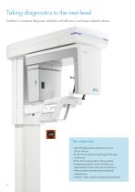
■ Ideal 3D imaging volume matched to the jaw arch (0 130 x 85 mm) ■ 0 130 x 50 mm volumes for single images of the upper and lower jaw ■ Excellent image quality in 2D and 3D thanks to the high-resolution Csl sensor with a pixel size of 49.5 pm ■ Reduced radiation dose thanks to the anatomically adapted volume ■ VistaSoft - modern, ergonomic image processing software
Open the catalog to page 2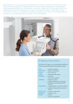
Ideal imaging volume, easy positioning, high image quality: VistaVox S represents a milestone in the field of 3D X-ray systems. Thanks to its unique technology, the 3D images generated with this system cover everything you need for reliable diagnostics, well-founded treatment decisions and convincing patient communication. In addition, the S-Pan technology of VistaVox S also enables pinpoint-accurate OPG image acquisitions in superior Durr Dental quality. Thanks to a high-resolution Csl sensor with a pixel size of 49.5 pm, you can benefit from exceptional image quality - both in 3D and in 2D....
Open the catalog to page 3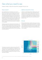
See what you need to see VistaVox S offers an ideal 3D volume that is adapted to the jaw arch Almost universal fit The jaw-shaped field of view of the VistaVox S shows an area of In addition to jaw-shaped images and the two additional volume sizes for Ø 130 x 85 mm volume that is relevant for diagnostics and is thus visibly the upper and lower jaw, VistaVox S offers ten further Ø 50 x 50 mm larger than many common volumes. The advantage of this is that by volumes: five each for the upper jaw and for the lower jaw. These are used changing the volume shape, VistaVox S also shows the rear molar...
Open the catalog to page 4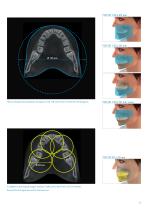
The jaw-shaped volume displays the region of a Ø 130 volume that is relevant for the diagnosis. In addition to jaw-shaped images, VistaVox S offers ten further Ø 50 x 50 mm volumes: five each for the upper jaw and for the lower jaw
Open the catalog to page 5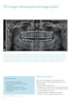
VistaVox S offers not only excellent value for money, but will also help you and your surgery team to increase your flexibility. In addition to CBCT images, you can also use VistaVox S to generate brilliant OPGs, which set new standards in the image sharpness of extraoral images. Thanks to this versatility, the new VistaVox S will really add value to your surgery. The unit also raises the bar in terms of efficiency. It enables the scan of a complete OPG image in a very short time of just seven seconds with an exceptionally low radiation dose. This will save you valuable time in comparison to...
Open the catalog to page 6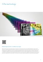
S-Pan technology Reliable diagnoses thanks to incredibly sharp images With S-Pan technology, the image regions that best correspond to the actual patient anatomy are automatically selected from a large number of parallel layers. These image parts are merged to form a panoramic image, which focuses on the actual anatomy of the patient. Deviations from the “average dentition” are taken into account, as are individually-angled teeth. The result is an image of impressive clarity, in which you will be able to immediately and effortlessly locate all anatomically relevant structures. Since the reconstruction...
Open the catalog to page 7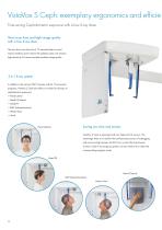
VistaVox S Ceph: exemplary ergonomics and efficie Time-saving Cephalometric exposure with a low X-ray dose Short scan time and high image quality with a low X-ray dose The very short scan time of just 1.9 seconds helps to avoid motion artefacts and to reduce the radiation dose. The modern high-sensitivity CsI sensors enable excellent image quality. 3-in-1 X-ray system In addition to the various CBCT volumes and the 17 panoramic programs, VistaVox S Ceph also offers six modes for all types of cephalometric exposures: ▪ Head Lateral ▪ Head Full Lateral ▪ Head PA ▪ SMV (submentovertex) ▪ Waters...
Open the catalog to page 8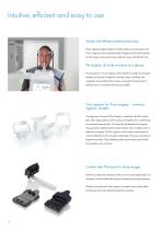
Intuitive, efficient and easy to use Simple and efficient patient positioning Three alignment lights (Sagittal, Frankfurt plane and Canine) for 2D X-ray images and two alignment lights (Sagittal and Frankfurt plane) for 3D images make positioning a pleasant, easy and efficient task. The display: all of the functions at a glance The innovative 7“ touch-display of the VistaVox S guides the therapist reliably and clearly through the necessary steps. Handling and navigation are exceptionally intuitive, ensuring that the process of taking X-rays is as smooth and easy as possible. Chin supports for...
Open the catalog to page 10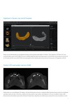
Digitalisation of plaster casts and drill templates Plaster casts and drill templates can be digitalised with the aid of the optional model holder for VistaVox S. The VistaSoft tool “DICOM-to-STL” then converts the data to open source STL files for use e.g. In VistaSoft Implant & Guide for planning purposes or so that the data can be printed out with the aid of a 3D printer. Both the scans and the STL files are saved together with the relevant patient to ensure that you have access to all image data at all times. VistaVox S 3D metal artefact reduction (MAR) Metal artefacts are a real challenge...
Open the catalog to page 11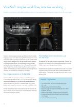
VistaSoft: simple workflow, intuitive working VistaSoft is a particularly comfortable and efficient solution for the capture, editing and display of digital 2D and 3D X-ray images. VistaSoft is intuitive to operate and opens up additional options for reliable diagnostics. Images can be edited using digital filters that adjust the contrast Automated panoramic reconstruction is just one click away and sharpness of the X-ray image to assist the diagnosis. The network-capable software supports the export of DICOM data as well as various interfaces The rendered OPG view makes it easier to navigate...
Open the catalog to page 12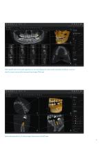
With VistaSoft and its AI-assisted algorithms you can easily display the automatically calculated mandibular canal and check its correct course via the transversal layer images (TSA view). Implant planning with a 5 x 5 volume image. Shown here in the MPR view. 13
Open the catalog to page 13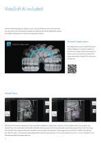
With the VistaSoft package you will gain access to powerful AI features that will not only make your day-to-day work in the practice noticeably more effective, but will also significantly increase the reliability of diagnostic work ahead of complicated procedures. Automatic image rotation The imaging software uses an algorithm based on artificial intelligence to check the orientation of intraoral X-ray images with the aid of anatomical features shown and corrects the rotation of the image automatically if needed. This will save you valuable time. VistaSoft Trace With the aid of innovative and...
Open the catalog to page 14All Dürr Dental catalogs and technical brochures
 lunos
7 Pages
 Product_catalogue_2026
216 Pages
 Tornado Compressors
5 Pages

Archived catalogs
 VistaSystem
16 Pages
 Dosingplan
2 Pages
 DürrConnect
32 Pages
 OroCup
4 Pages
 VC 45
4 Pages
 Brochure Compressors
12 Pages
 Power Tower Silence 200
1 Page
 dsh
11 Pages
 VistaVox S
12 Pages
 VistaCam iX
6 Pages
 Hygoclave 90
12 Pages
 Laboratory
8 Pages
 Brochure DBSWIN 5
6 Pages
 PTS 200
2 Pages
 Brochure Vector
12 Pages
 V 2400
2 Pages
 Hygiene-Fibel
20 Pages
 Vista Pano S Ceph
12 Pages
 Tyscor Pulse
2 Pages
 AR Marker
1 Page
 Brochure VistaScan Mini Easy
2 Pages
 CA 4
2 Pages
 Hygoclave 30
4 Pages
 Tyscor V 2
10 Pages
 Brochure Tyscor Pulse
6 Pages
 Brochure Clinics
10 Pages
 V/VS suction system
18 Pages
 Hygopac
2 Pages
 Cannula brochure
6 Pages
 Brochure System-Hygiene
16 Pages
 DSH range package
4 Pages
 hygiene technology
14 Pages
 PTS 120
4 Pages
 Periomat Plus
6 Pages
 Brochure digital Xray
12 Pages
 VistaPano S
8 Pages
 Vector Scaler
4 Pages
 Duo Tandem Med
2 Pages
 Brochure VistaIntra DC
4 Pages
 V / VS / VSA 300 S
18 Pages
 CA 1
12 Pages
 General catalog
24 Pages
 Brochure Imaging
10 Pages
 Vector Paro
6 Pages
 Tornado
8 Pages
 Film developing
2 Pages


- Sterilizer
- Steam sterilizer
- Analysis software
- Automatic sterilizer
- Stainless steel sterilizer
- Benchtop sterilizer
- Cannula
- Dry compressor
- Radiology software
- Hospital compressor
- Visualization software
- Flat panel detector
- Dental compressor
- Reporting software
- Gas filter
- Camera with USB port
- Sterilizer with touchscreen
- Programmable sterilizer
- Planning software
- Cloud-based software